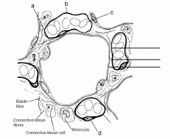
38 on the diagram below identify alveolar epithelium
The epithelium here remains low cuboidal. Each respiratory bronchiole branches into between 2 and 11 alveolar ducts that still contain smooth muscle fibers in their walls. Along these walls, the alveolar ducts give rise to single alveoli and to numerous alveolar sacs, which are associated with 2 to 4 alveoli. Aug 12, 1996 — Trachea, identified by the presence of hyaline cartilage in its wall. ... Notice how cellular the c.t. under the epithelial layer is.
On the diagram below identify alveolar epithelium capillary. Fill in the blanks with the terms provided. Appropriately label all structure provided with leader lines on the diagrams below. Bronchials trace a molecule of oxygen from the nostrils to the pulmonary capillaries of the lunges. Appropriately label all structures provided with leader ...

On the diagram below identify alveolar epithelium
On the diagram below, identify alveolar epithelium, capillary endothelium, alveoli, and red bloo... A: Lungs provide the respiratory surface for the exchange of gases. Alveoli are very small sacs present... 18.11.2021 · A comprehensive database of more than 35 histology quizzes online, test your knowledge with histology quiz questions. Our online histology trivia quizzes can be adapted to suit your requirements for taking some of the top histology quizzes. The lungs are located in the chest on either side of the heart in the rib cage.They are conical in shape with a narrow rounded apex at the top, and a broad concave base that rests on the convex surface of the diaphragm. The apex of the lung extends into the root of the neck, reaching shortly above the level of the sternal end of the first rib.
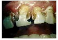
On the diagram below identify alveolar epithelium. Start studying Lab 3 (exercises 5,6,7). Learn vocabulary, terms, and more with flashcards, games, and other study tools. The alveolar wall or septum is made up of three tissue components: surface epithelium, supporting tissue, and an extensive network of continuous capillaries. Centrally it has capillaries surrounded by a vibrant network of elastin, reticular, and collagen fibers with a layer of squamous epithelial of two adjacent alveoli on either side. On the diagram below, identify the alveolar duet, respiratory bronchioles, terminal bronchiole, alveoli, and alveolar sa. Examining Prepared Slides of Tracheal and Lung Tissue 14. The tracheal epithelium is ciliated and has goblet cells. What is the function of each of these modifications? cilia: goblet cells: 15. Dental anatomy is a field of anatomy dedicated to the study of tooth structure. The development, appearance, and classification of teeth fall within its field of study, though dental occlusion, or contact between teeth, does not.Dental anatomy is also a taxonomic science as it is concerned with the naming of teeth and their structures. This information serves a practical purpose for dentists ...
B. Alveolar ducts The walls of alveolar ducts View Image are lined by alveoli and alveolar sacs (clusters of alveoli). C. Alveolus The walls of these structures are covered on both sides by squamous epithelium (too thin to see) of Type I cells lining adjacent alveolar lumens. Within the walls is an extensive capillary network. Underneath the thin skin of the nose are its skeletal features. ... An olfactory epithelium used to detect odors is found deeper in the nasal cavity. parts, epithelial tissues form the inner lining and external lining of body parts. To summarize, the apical pole faces the surface, while the basal pole is attached to the connective tissue located below the epithelium. Types of epithelial tissue. There are 3 different types of epithelial tissue: squamous, cuboidal, and columnar. From these data, the authors suggested that stretching of alveolar epithelial cells is of high relevance above 80% of TLC while below 80% TLC, deformations without much change in the surface area of the basal lamina (and, therefore, stretch of alveolar epithelial cells) dominate micromechanics, e.g. unfolding/folding of septal walls or changes ...
The simple epithelial tissue is a closed network of flat epithelial cells. These are located on the basal membrane. It is composed of a single layer of cells that are specialized in diffusion, osmosis, filtration, secretion, and absorption.The simple epithelial tissue is found in the alveolar epithelium (pulmonary alveolus), the endothelium (lining of blood vessels and lymph vessels), and the ... Simple squamous epithelium. Simple squamous epithelia consist of a single layer of flattened cells. This type of epithelia lines the inner surface of all blood vessels (endothelium), forms the wall of alveolar sacs in the lung and lines the body cavities (mesothelium). PDL, alveolar bone, junctional epithelium ... Diagram of the reciprocal induction of odontoblasts and ameloblasts. ... they put pressure on bone tissue in the alveolar sockets below. Pressure on bone tissue triggers release of BMPs which leads to the deposition of bone tissue, pushing teeth outwards. by N Kia'i · 2021 · Cited by 12 — Humidification requires serous and mucous secretions, and warming relies on the extensive capillary network that lays within the alveoli. The ...
Page from a Manuscript with Diagrams Protecting against Children's Illnesses (19th century) // Tibet
On the diagram below identify alveolar epithelium capillary. Leading Edje Home Bronchials trace a molecule of oxygen from the nostrils to the pulmonary capillaries of the lunges. Appropriately label all structures provided with leader lines on the diagrams below. Back to notecard set easy notecards home page.
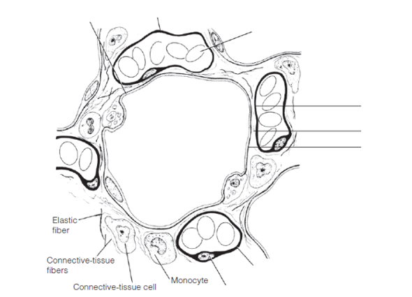
On the diagram below, identify alveolar epithelium, cap ...
- The epithelial lining of alveoli consists mainly of type I alveolar cells (also known as type I pneumocytes). - These are large, flat, squamous cells with few organelles and thin cytoplasm. - They cover about 93% of alveolar surface area. - Their primary purpose is air-blood gas exchange. - The junctions between these cells are narrow (1nm).
Expression of MT I+II proteins in the alveolar epithelium ...
The epithelium of the nasal passages, for example, is essential to sensing odors, and the bronchial epithelium that lines the lungs can metabolize some airborne carcinogens (Betts, et al., 2013). the respiratory zone includes structures that are directly involved in gas exchange (Betts, et al., 2013)

Respiratory System page 21
Solution for 14. On the diagram below, identify alveolar epithelium, capillary endothelium, alveoli, and red blood cells. Bracket the respiratory membrane.…

burger and fries on brown wooden round plate
The intraalveolar septa are the partition of two adjacent alveoli. They consists of two thin squamous epithelium layer. Mucosa layer of alveoli. Alveolar duct and alveolar sac lined by the patches of simple cuboidal epithelium. But alveolus is lined by simple squamous epithelium. These simple epithelium of alveoli contain the three major cells ...

Injury of alveolar epithelial cells. Normal alveolar ...
The alveolar epithelium consists primarily of simple squamous epithelium. The squamous epithelial cells, called pneumocytes _____, are unusually thin and delicate. type 2. Pneumocytes ____, also called septal cells, are scattered among the squamous cells. surfactant.

gray rope on brown wooden table
Question: iagram below, identify alveolar epithelium, capillary endothelium, alveoli, and red blood cells. Bracket the brane respira Elastic fiber Connective tissue fibers Monocyte . This problem has been solved! See the answer See the answer See the answer done loading. Show transcribed image text

gray concrete statue of a man
1 день назад · For example, transplantation of either Tgfb1-/-epithelium 73 or Tgfb1+/-epithelium, which has dramatically reduced TGFB1 levels due to the absence of positive feedback 72, into WT mammary fat pads results in increased epithelial proliferation and ductal extension, while overexpression of dominant negative TGF beta receptor II (TGFBR2) in the epithelium leads to alveolar hyperplasia 76.

Observe the figure given below. Identify the marking 'X ...
R.J. Mason, in Encyclopedia of Respiratory Medicine, 2006 The alveolar epithelium is composed of two types of epithelial cells, named alveolar type I and type II cells. Type I cells are large flat cells that comprise about 95% of the alveolar surface. Type II cells are small cuboidal cells with characteristic lamellar inclusions and apical microvilli and cover about 5% of the alveolar surface.

Mobile Delousing Unit, Truck Equipment, Plan of Operation and Erection Procedure, Presentation Drawing (1943) // Bertrand Goldberg American, 1913-1997
Alveolar sac - 2 or more alveoli sharing a common opening 2 types of alveolar epithelial cells Type I alveolar cells - form nearly continuous lining, more numerous than type II, main site of gas exchange Type II alveolar cells (septal cells) - free surfaces contain microvilli, secrete alveolar fluid (surfactant
white animal skull on white surface
The diagram below shows the oxygen levels (paO: 2) in the alveoli and at ... Using only the letters A to G from the diagram, identify the following (letters may be used ... the epithelial cells covering the villi of a snake from each of Groups 1 and 2. microvilli 0.5 μm

Fangruida: New coronavirus research 2020v4.0 electronic version (Davis.k)
Primary human alveolar epithelial cells (hAECs) were populated on the surfaces of the sacs to form the monolayer epithelium. The alveolar lung model was further subjected to cyclic inhalation/exhalation movements, as well as used to investigate the effects of cigarette smoke and severe acute respiratory syndrome coronavirus 2 (SARS-CoV-2 ...

cooked food on white ceramic plate
In summary, the following things can be said about the alveolar shape and structure: Largely polyhedral shape. Open at one end, like a cup. Walls of the alveoli are composed of the pulmonary capillary sheet. Alveolar surfaces are covered in a thin (200nm) layer of surfactant which acts as the interface with the gas.

sliced orange fruit on brown round woven round bowl
The alveolar epithelium lines the wall of the alveolus by the creating an ALI that facilitates an exchange of gases and is estimated to cover an average surface area of 100-140 m 2 in humans. 102 The alveolar epithelium is made up of two epithelial cell types, namely, the terminally differentiated squamous alveolar epithelial type-1 (AT-I) and the surfactant-producing cuboidal alveolar ...

Drachm (Coin) Depicting a Cow with Dolphin below (about 416-357 BCE) // Greek
The epithelium of the alveoli, contains two main types of cells: type I pneumocytes: large flattened cells - (95% of the total alveolar area) which present a very thin diffusion barrier for gases. type II pneumocytes (making up 5% of the total alveolar area, but 60% of cells). These cells secrete 'surfactant' which decreases the surface tension ...
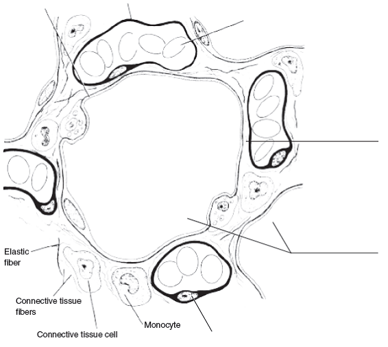
On the diagram below, identify alveolar epithelium, cap ...
Start studying A&P2 Lab 13 HW, A&P2 Lab 12 HW, A&P2 Lab 11 HW, A&P2 Lab 10 HW, Lab 9 HW, Lab 8 HW, A&P2 Lab 1 HW, A&P2 Lab 2 HW, A&P2 Lab 3 HW, A&P2 Lab 4 HW, A&P 2 Lab 5 HW, A&P2 Lab 6 HW, A&P2 Lab 7 HW. Learn vocabulary, terms, and …

burger on brown wooden table
The alveolar region is a branching system of alveolar ducts whose walls are made of alveoli. The alveolar ducts end into alveolar sacs which contain alveolar outpouches. The alveoli thus forms the walls of alveolar ducts and sacs.The openings or the mouths of alveoli consist of a dense network of elastic and collagen fibers.

Pulmonary alveolus - wikidoc
Types of Epithelial Tissue. There are three types of epithelial cells, which differ in their shape and function. Squamous- thin and flat cells Cuboidal- short cylindrical cells, which appear hexagonal in cross-section Columnar- long or column-like cylindrical cells, which have nucleus present at the base On the basis of the number of layers present, epithelial tissue is divided into the simple ...

Microanatomy of the Lung - Respiratory - Medbullets Step 1
On the diagram below identify aveolar epithelium, capillary endothelium, alveoli, land red blood cells. Bracket the respiratory membrane. Elastic.4 pages

Ultrastructure of alveolar epithelial Type I cells 24 ...
Respiratory epithelial cells line the respiratory tract from trachea to bronchi into bronchioles and alveolar sacs. The primary functions of the respiratory ...Missing: diagram identify
Alveoli: Anatomy, function and clinical points | Kenhub
Ciliated columnar epithelium is composed of simple columnar epithelial cells with cilia on their apical surfaces. These epithelial cells are found in the lining of the fallopian tubes where the assist in the passage of the egg, and parts of the respiratory system, where the beating of the cilia helps remove particulate matter.

Sр-В expression in alveolar epithelium, that covering ...
On the diagram below identify alveolar epithelium capillary. Appropriately label all structures provided with leader lines on the diagrams below. Label all structures provided with leader lines on the diagram below. Fill in the blanks with the terms provided. Plveoll superior lobe middle lobe inferior lobe.

sliced orange fruit in brown woven round basket
Identify the tissue type and its function. Simple Squamous Epithelium •Diffusion and Filtration •Secretes lubricating substances in serosae. Identify the structure indicated. Skeletal Muscle. MultiplePeripherally Located Nuclei. Identify the structure indicated. Neuron Cell Body .
Lab 9 Quiz Study Flashcards - Cram.com
14. On the diagram below, identify alveolar epithelium, capillary endothelium, alveoli, and red blood cells. Bracket the respiratory membrane. VÉeu É Elastic fiber O st W Connective-tissue fibers I Monocyte Connective-tissue cell 15. Why does oxygen move from the alveoli into the pulmonary capillary blood? CIS 770 16.

Marina City: Finish Diagram (n.d.) // Bertrand Goldberg American, 1913-1997
The diagram given below is a simple epithelium. (MARCH-2015) a) Name the part marked as "P" in the figure. b) Write one function of a simple epithelium. Answer: a) Cell membrane (Out of syllabus) b) They are found in the walls of blood vessels and air sacs of lungs and are involved in a function diffusion. Question 21.

Dymaxion Car, Section (1933) // Richard Buckminster Fuller American, 1895-1983
Histological slides illustrating type I pneumocytes (left) and type II pnemocytes (right) Type II pneumocytes. The type II alveolar cells (also known as type II pneumocytes) have two functions: (1) to repair the alveolar epithelium when squamous cells are damaged, and (2) to secrete pulmonary surfactant. Surfactant is composed of phospholipids and protein, and coats the alveoli and smallest ...
Human Structure Virtual Microscopy
The lungs are located in the chest on either side of the heart in the rib cage.They are conical in shape with a narrow rounded apex at the top, and a broad concave base that rests on the convex surface of the diaphragm. The apex of the lung extends into the root of the neck, reaching shortly above the level of the sternal end of the first rib.

Lung immaturity with cuboidal alveolar epithelium in a ...
18.11.2021 · A comprehensive database of more than 35 histology quizzes online, test your knowledge with histology quiz questions. Our online histology trivia quizzes can be adapted to suit your requirements for taking some of the top histology quizzes.

Pulm Formative Review Flashcards | Quizlet
On the diagram below, identify alveolar epithelium, capillary endothelium, alveoli, and red bloo... A: Lungs provide the respiratory surface for the exchange of gases. Alveoli are very small sacs present...
Dysfunction of microvascular endothelium and alveolar ...

Diagram of mammary alveolus and alveolar epithelial cell ...

Fangruida: New coronavirus research 2020v4.0 electronic version (Davis.k)

white ceramic bowl with green leaves

Measuring Type 1 alveolar epithelial cell ratio. The Type ...

Club de Centre Rural: Perspective Sketch (1943) // Le Corbusier French, born Switzerland, 1887-1965

white spiral paper on black surface

Alveolar space in ALI/ARDS. Injury of alveolar epithelial ...
white and yellow ice cream with cone
0 Response to "38 on the diagram below identify alveolar epithelium"
Post a Comment