38 gram negative bacteria diagram
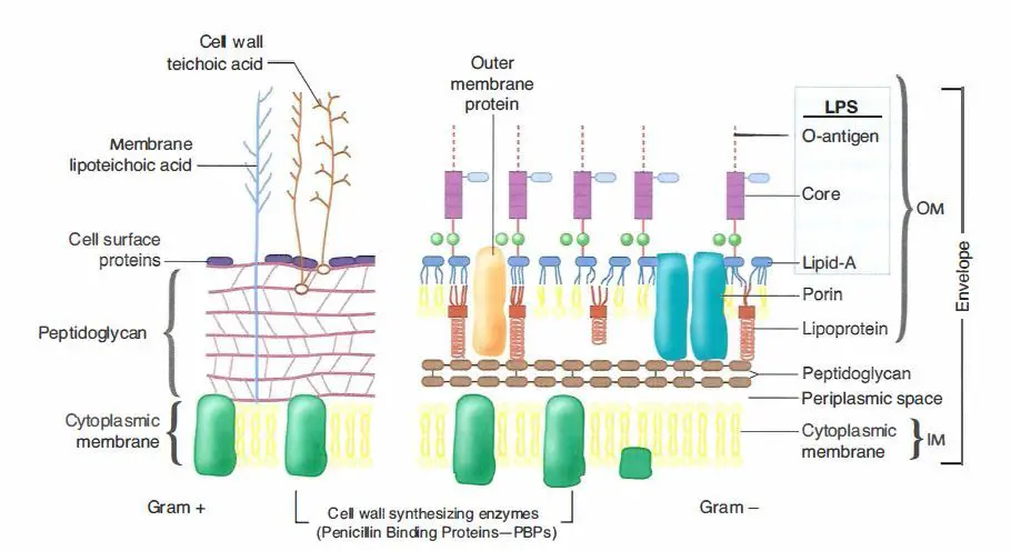
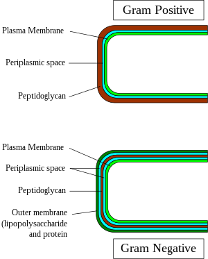
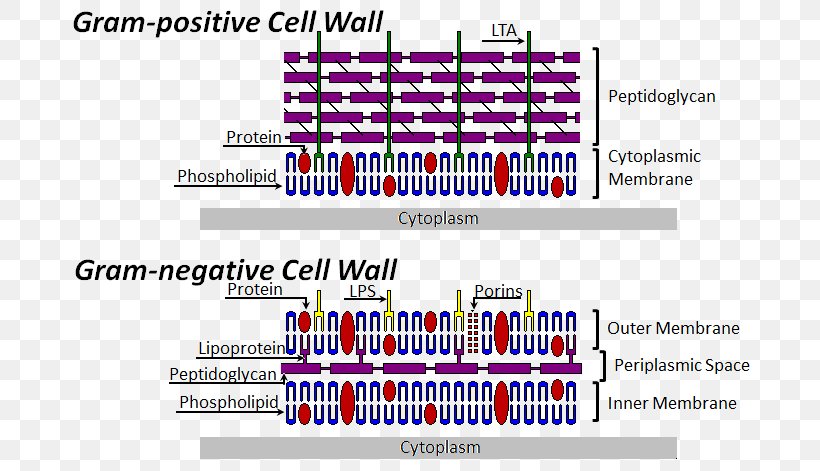
After lysozyme digestion gram negative bacteria become spheroplast. 20. Susceptibility to penicillin and sulphonamide. Usually cocci and rod shaped spore forming bacteria (exception Corynebacterium and Lactobacillus are non-spore forming rod). 'Gram-positive' and 'gram-negative' are terms used to broadly categorize two different types of bacteria. The cell walls of gram-negative bacteria contain only a thin layer of peptidoglycan, but they also have an outer membrane that is absent in gram-positive bacteria.
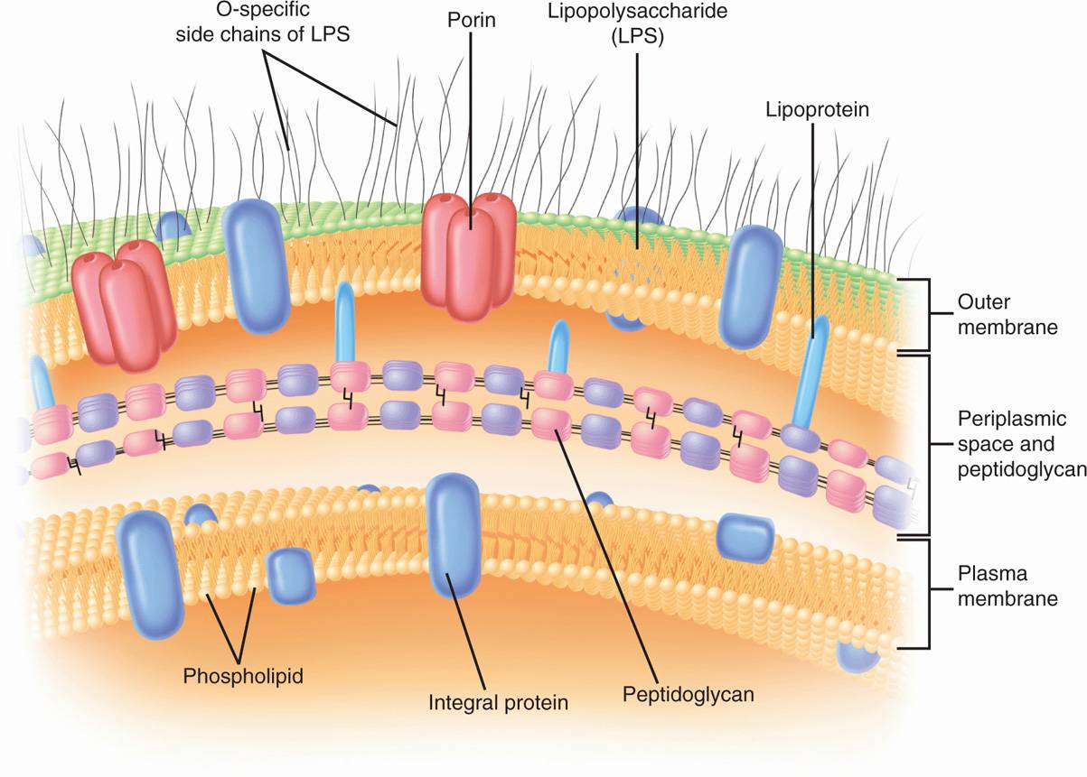
Apr 11, 2021 · Gram-negative bacteria cell wall. The Cell wall of the Gram-Negative Bacteria is very complex as compared to that of Gram-Positive Bacteria. Combined with the major role of the outer membrane of the cell, with a layer of peptidoglycan, its functional properties are complex, and here is a description of the cell wall and its functional parts.

Gram negative bacteria diagram
8. According to Peberdy (1980) the only compound present in the cell walls of both Gram-negative and Gram-positive bacteria is ‘peptidoglycan’. The cell walls of Gram-positive bacteria contain up to 95% peptidoglycan and up to 10% teichoic acids. 9. Cytoplasmic membrane is a thin (5-10 nm) layer lining the inner surface of the cell wall. Gram-negative bacteria are found everywhere, in virtually all environments on Earth that support life. The gram-negative bacteria include the model organism Escherichia coli, as well as many pathogenic bacteria, such as Pseudomonas aeruginosa, Neisseria gonorrhoeae, Chlamydia... Gram Negative Bacteria Schema Demonstrated by: Yomna Hagag Recorded by: Muhammad Aladawi Under Supervision of: Dr.Rasha Brawa Head of Microbiology...
Gram negative bacteria diagram. Gram-negative bacteria have higher levels of transport proteins, which remove toxic substances, such as antibiotics from the cells. Certain gram-negative bacteria can also acquire antibiotic resistance via mutation or acquisition of foreign DNA through gene transfer. One well-known example of this is E... Gram negative bacteria tend to employ small molecules as auto-inducers of master regulators of gene expression, while gram positive bacteria favor oligopeptides. Gram-negative bacteria are inherently resistant to macrolide antibiotics, presumably due to the sizes of the molecules (molecular weights of... May 17, 2021 · Bacteria can be classified based on shape, mode of nutrition, respiration, the composition of the cell wall, etc. Based on these criteria, bacteria can be classified as bacillus, coccus or vibrio, etc., autotrophic or heterotrophic, aerobic or anaerobic, Gram + or Gram -. susceptibilitytoantibiotics.Antibioticresistanceofthegramnegativebacteriaisgivenbytheoutermembranepresent. inthesebacteria.Neisseriagonorrhoeae,PseudomonasaeruginosaandYersiniapestis...
Bacteria are a commonly used organism for studies of genetic material in the research laboratory. ... which diagram of a cell wall is a gram-negative cell wall? A) a B) b Gram positive bacteria appear purple and gram-negative bacteria appear pink when stained by Gram-staining methods. Gram-negative bacteria contain an extra layer of cells called outer membrane or lipopolysaccharide (LPS) layer which surrounds the thin peptidoglycan layer. Gram negative, kidney bean-shaped diplococci. Oxidase and catalase positive. Large polysaccharide capsule (latex particle agglutination to identify capsular antigens in CSF). Contains Vancomycin to prevent growth of gram positive bacteria, polimyxin to prevent growth of other gram negative... Gram-negative bacteria are bacteria that do not retain the crystal violet stain used in the gram-staining method of bacterial differentiation. They are characterized by their cell envelopes, which are composed of a thin peptidoglycan cell wall sandwiched between an inner cytoplasmic cell...
Like Gram positive bacteria, Gram negative bacteria are also well distributed in different environments across the globe. · Gram negative bacilli - There are different groups of Gram negative bacilli bacteria including those described as Anaerobic or Aerobic. **Other discussions** [Episode 1 - Pneumococcus](https://reddit.com/r/anime/comments/8z6vpb/hataraku_saibou_ep_1_doctors_notes/) [Episode 2 - Scrape wound](https://www.reddit.com/r/anime/comments/90bxnl/hataraku_saibou_ep_2_doctors_notes/) [Episode 3 - Influenza](https://reddit.com/r/anime/comments/913mov/hataraku_saibou_ep_3_doctors_notes/) [Episode 4 - Food poisoning](https://reddit.com/r/anime/comments/93a8xt/hataraku_saibou_ep_4_doctors_notes/) [Episode 5 - Cedar pollen allergy](https:/... Gram-negative bacteria refers to a broad category of bacteria that are unable to retain the crystal violet dye owing to their distinct cell wall structure. Know more about such bacteria with respect to their cell wall structure, examples, infections and treatment options. Gram negative bacteria are later stained with safranin or fuchsin for observation under microscope. Gram negative bacteria after safranin or fuchsin staining will appear red or pink colour. Gram staining differentiates bacteria by the chemical and physical properties of their cell walls by detecting the...

Blue and white texture with negative space > > > If you interested in my artwork, please visit and follow instagram.com/flyd2069
Gram-negative and gram-positive bacteria stain differently because their cell walls are different. They also cause different types of infections, and Gram-negative bacteria are enclosed in a protective capsule. This capsule helps prevent white blood cells (which fight infection) from ingesting the bacteria.
What's the difference between Gram-negative Bacteria and Gram-positive Bacteria? Danish scientist Hans Christian Gram devised a method to differentiate two Gram-negative bacteria are also more resistant to antibiotics because their outer membrane comprises a complex lipopolysaccharide (LPS)...
"Gram negative coccobacilli" may suggest Haemophilus species. "Lactose-positive gram negative rods" may suggest Enterobacteriaceae, such as E. coli, Klebsiella, or Enterobacter spp.
Low (acid-fast bacteria have lipids linked to peptidoglycan). High (because of presence of outer membrane). 11. the difference is clear but in simple explanation gram staining is what makes bacteria to be gram positive or negative and this happens because gram positive bacteria have...
Hey guys! So, I’ve been reading about horseshoe crabs for fun and I had some questions. Firstly, are there different type of endotoxins? To my understanding endotoxins are LPS (lipopolysaccharide) found only on gram negative bacteria. So why do we use LAL if we can use the gram staining method to detect between Gram - and gram + ? Can we detect endotoxins using gram staining..? Secondly, if there is an alternative to LAL called Recombinant factor C (rFC) assay how come some pharmaceutical c...
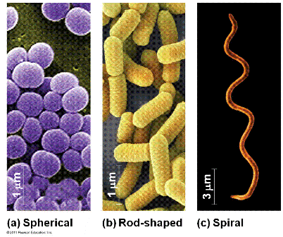
Gram negative coccobacillus bacteria have bacterial shapes that are in between spherical and rod shaped. Bacteria of the genus Haemophilus and Acinetobacter are coccobacilli that cause serious infections. Haemophilus influenzae can cause meningitis, sinus infections, and pneumonia.
Gram-negative bacteria are bacteria that do not retain the crystal violet stain used in the gram-staining method of bacterial differentiation.
Gram negative bacteria are composed of a cell envelope in the outside of the cell wall, called the outer membrane, which is 7.5-10 nm thick. Gram Positive Bacteria: Gram positive bacteria retain the crystal violet stain during gram staining, giving the positive result.
“Research has suggested that the etiology of gastroesophageal reflux disease-related esophagitis includes a cytokine-mediated inflammatory component and is, therefore, not merely the result of esophageal mucosal exposure to corrosives (i.e., acid).” “We discuss the potential role for manipulating the EM (esophageal mucosa) as a therapeutic option for treating the root cause of various esophageal disease rather than just providing symptomatic relief (i.e., acid suppression).” “gram-negative pro...
First Described: Gram-negative bacteria were first described in Berlin by the Danish scientist Hans Christian Gram in 1884 when he used Gram Gram-negative bacteria are widespread causes of opportunistic infections in dogs and cats and primarily cause disease when host defenses are impaired.
The treatment of multidrug-resistant Gram-negative bacteria (MDR-GNB) infections in critically ill patients presents many challenges. Since an effective treatment should be administered as soon as possible, resistance to many antimicrobial classes almost invariably reduces the probability of...
Gram-positive and Gram-negative bacteria are classified based on the ability to retain the gram stain. The gram-positive bacteria would retain the gram stain and observed as violet color after the application of iodine (as mordant) and alcohol (Et...
Bacteria can either be a gram-positive or gram-negative, and to find it out, a gram-staining technique has to be used. Gram Staining technique is the most Along with their staining characteristics, Gram Positive and Gram Negative bacteria differ from each other in various aspects which are listed below
Gram negative bacteria appear a pale reddish color when observed under a light microscope following Gram staining. This is because the structure of their cell wall is unable to retain the crystal violet stain so are colored only by the safranin counterstain. Examples of Gram negative bacteria include...
It consists of lipopolysaccharides, lipids and proteins. The outer membrane has hydrophilic channels of 16-stranded (3-barrel proteins called porins. The single layered cell wall of Gram positive bacteria and inner wall layer of Gram negative is made up of pepidoglycan, proteins, non-cellulosic carbohydrates, lipids, amino acids, etc.
Gram positive bacteria stain blue-purple and Gram negative bacteria stain red. The difference between the two groups is believed to be due to a much larger peptidoglycan (cell wall) in Gram positives. As a result the iodine and crystal violet precipitate in the.
Apr 15, 2021 · Gram-positive bacteria stain purple while the Gram-negative bacteria stain Pink, after losing the purple color during the alcohol was thus taking up the safranin. Gram staining has been especially used because of its ability to differentiate bacteria base on their cell wall content, a major characteristic that classifies bacteria into two types ...
Gram negative bacteria can be rod-shaped, round or spiral. They can be non-sporulating or sporulating. Gram negative bacteria stain pink during the process of Gram staining. In this article, we will describe what gram-negative bacteria are, define their structure and explore various...
Gram-negative bacteria do not retain the crystal violet stain and further, it takes the color of the red counterstain in Gram's method of staining. Answer: Gram-negative bacteria are considered more harmful. These cause certain diseases. Also, their outer membranes are hidden by a slime layer and it...
Photoautotrophic bacteria are Gram‐negative rods which obtain their energy from sunlight through the processes of photosynthesis. Chemoautotrophic (or chemolithotrophic) bacteria are a group of Gram‐negative bacteria deriving their energy from chemical reactions involving inorganic material.
Gram Negative Bacteria Schema Demonstrated by: Yomna Hagag Recorded by: Muhammad Aladawi Under Supervision of: Dr.Rasha Brawa Head of Microbiology...
Gram-negative bacteria are found everywhere, in virtually all environments on Earth that support life. The gram-negative bacteria include the model organism Escherichia coli, as well as many pathogenic bacteria, such as Pseudomonas aeruginosa, Neisseria gonorrhoeae, Chlamydia...
8. According to Peberdy (1980) the only compound present in the cell walls of both Gram-negative and Gram-positive bacteria is ‘peptidoglycan’. The cell walls of Gram-positive bacteria contain up to 95% peptidoglycan and up to 10% teichoic acids. 9. Cytoplasmic membrane is a thin (5-10 nm) layer lining the inner surface of the cell wall.












0 Response to "38 gram negative bacteria diagram"
Post a Comment