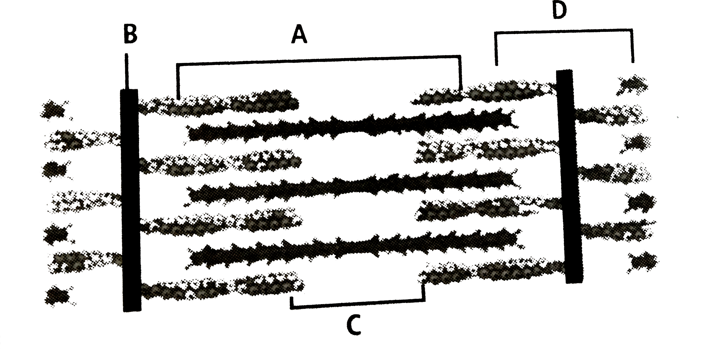
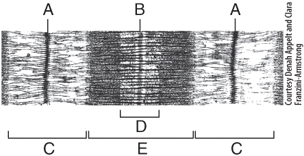
38 identify the structures labeled a, b, and c in the diagram of a sarcomere above.
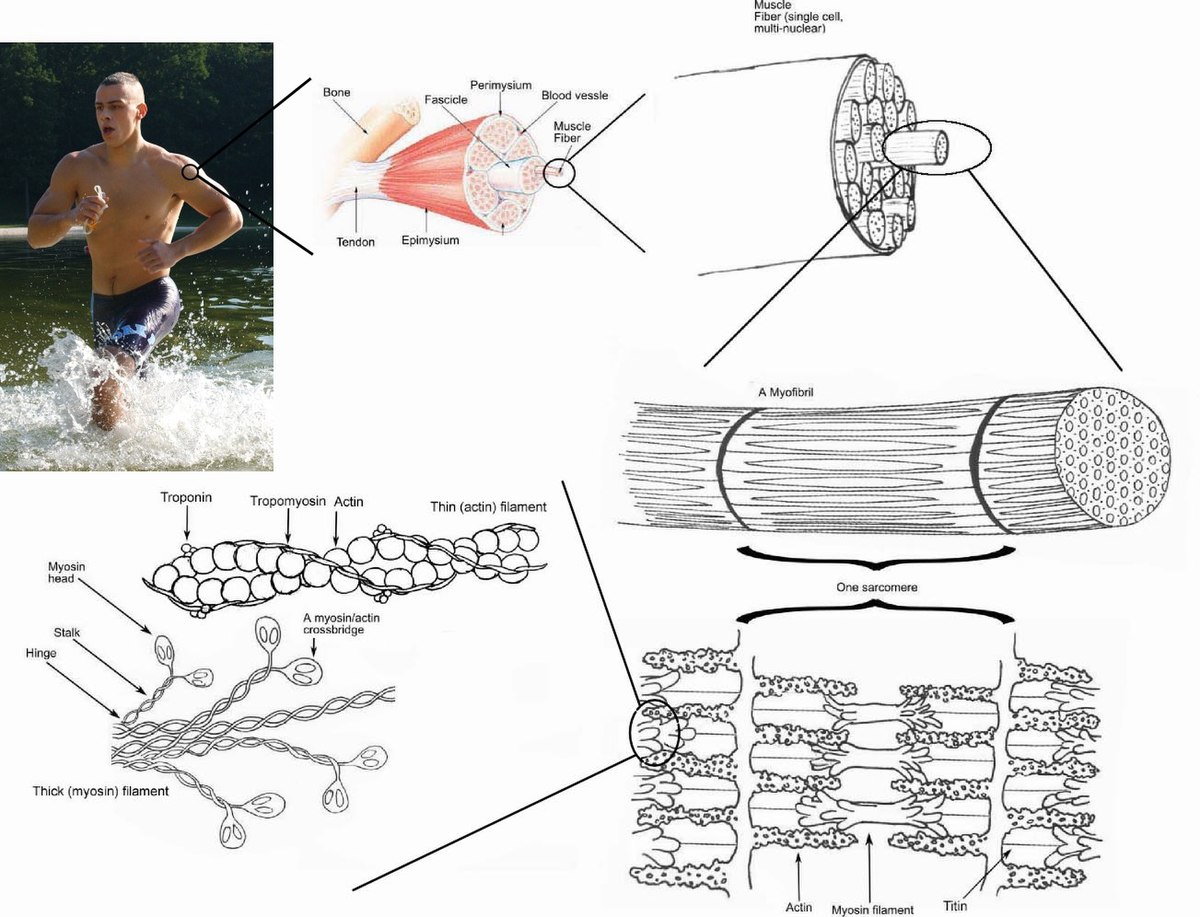
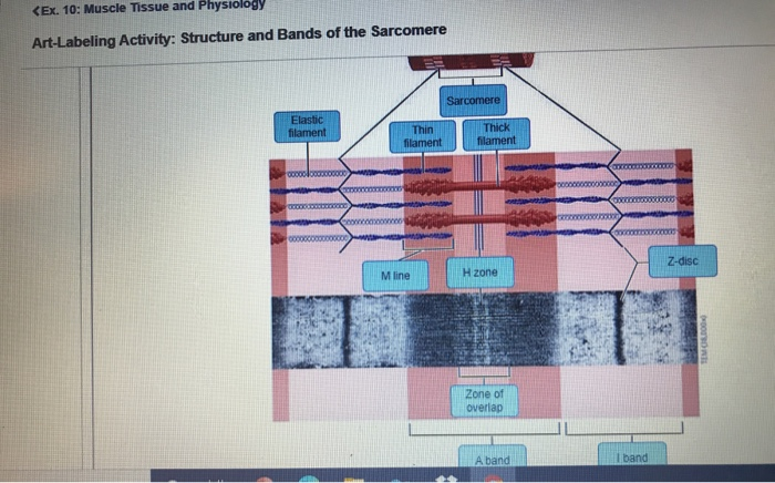
10.2 Skeletal Muscle - Anatomy & Physiology A sarcomere is defined as the region of a myofibril contained between two cytoskeletal structures called Z-discs (also called Z-lines), and the striated appearance of skeletal muscle fibers is due to the arrangement of the thick and thin myofilaments within each sarcomere (Figure 10.2.2). muscle worksheet.pdf - Muscle Contraction Prior ... - Course Hero A, B, and C above. 2. In the diagram below, draw three vertical lines showing the locations within a sarcomere of the cross sections indicated by Figures A, B, and C. Label each of the lines. A B thin thin thick thick C A B C
Animated Tutorial 33.1 Quiz Flashcards | Quizlet Identify the structures labeled A, B, and C in the diagram of a sarcomere above. A is a "blue" filament, attached to the Z line at the ends of a sarcomere. B is a "red" filament, crossing the M band. C is a "grey" filament attached to both B and the Z line.
Identify the structures labeled a, b, and c in the diagram of a sarcomere above.
Study 50 Terms | Chapter 10: Skeletal... Flashcards | Quizlet B) B C) C D) D E) E. A) A. Identify the letter that indicates the endomysium. A) A B) B C) C D) D E) E. ... connects a muscle to underlying structures through a flat sheet or web. B) consists of a neuron and all the muscle fibers it innervates. ... Which region of the sarcomere does not change in length during contraction? A) A band B) H zone C ... Labeled Sarcomere Diagram Jan 23, 2019 · Labeled Sarcomere Diagram. Their observations led to the discovery of sarcomere zones. Sarcomere The figure depicts the structure of a Sarcomere. (Each zone is labeled). They first. Start studying Sarcomere Labeling. Learn vocabulary, terms, and more with flashcards, games, and other study tools. BSC 2011 undergraduate Flashcards - Quizlet Identify the structures labeled A, B, and C in the diagram of a sarcomere above. A = Actin filament; B = Myosin filament; C = Titin The sliding filament theory states that during muscle contraction
Identify the structures labeled a, b, and c in the diagram of a sarcomere above.. PDF Muscle Contraction Model 1: Anatomy of a Sarcomere 11. There are three sarcomeres shown in the diagram below. Sarcomere 1 Sarcomere 2 Sarcomere 3 a) In Sarcomere 1, identify the location within the sarcomere of the cross section indicated by Figure A in Model 3. Draw a vertical line and label it A. b) In Sarcomere 2, identify the location within the sarcomere of the cross section Identify The Structures Labeled A B And C In The Diagram Of A ... In the diagram below draw three vertical lines showing the locations within a sarcomere of the cross sections indicated by figures a b and c. A b and c above. The diagrams in model 3 are cross sections of a sarcomere that show the filaments at various locations within a sarcomere. Play this quiz called label the sarcomere and show off your skills. pogil-muscle-contraction-wod document.docx - Model 2 ... Fig. A Fig. B Fig. C QUESTIONS: 11. Label the thick and thin filaments in Figs. A, B, and C above. 12. There are three sarcomeres shown in the diagram below. Sarcomere 1 Sarcomere 2 Sarcomere 3 a) In Sarcomere 1, identify the location within the sarcomere of the cross section indicated by Figure A in Model 3. Muscle Contraction - Science for the WINegar A, B, and C above. 2. In the diagram below, draw three vertical lines showing the locations within a sarcomere of the cross sections indicated by Figures A, B, and C. Label each of the lines. A B C thin thin thick thick A B C
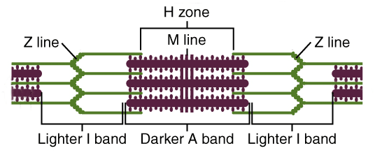
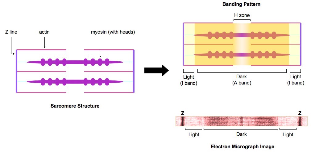
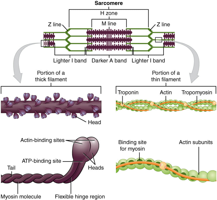
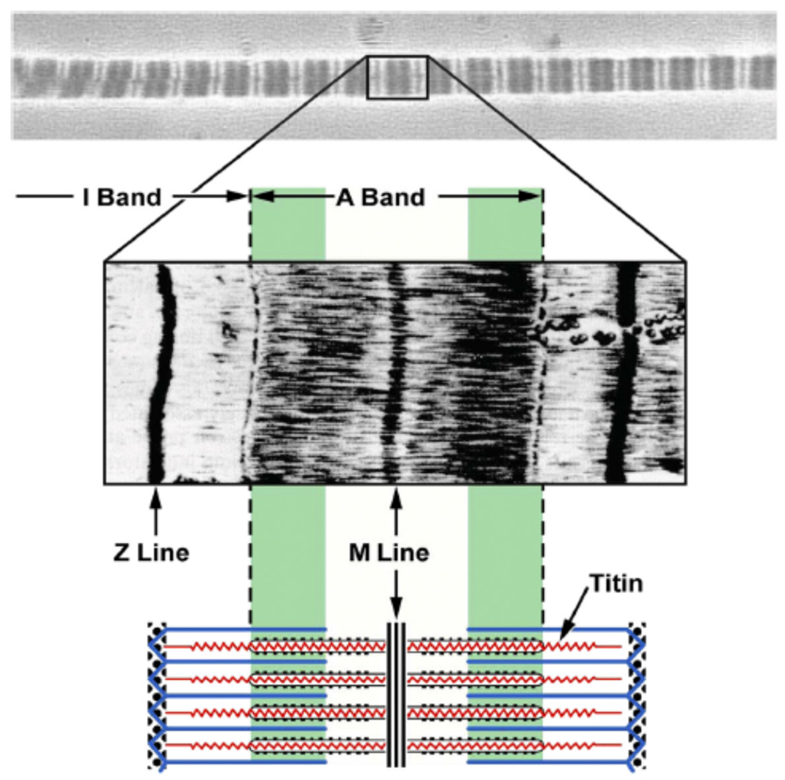
Sarcomere | Definition, Structure, & Sliding Filament Theory Sarcomere Definition. The sarcolemma or sarcomere is an excitable membrane cell and shares many properties with the cell membrane of the neuronal structure. The function of sarcolemma is to connect the basement membrane which wrapped all connective tissues. The sarcolemma also has an extracellular matrix containing various polysaccharides ... PDF ADVANCED MATRICULATION LEVEL 2021 SECOND SESSION Biology a. On Figure 1, clearly mark and label: i. using the letter A - the site of glycolysis; (1) ii. using the letter B - the site of Krebs' cycle; (1) iii. using the letter C - the site of the electron transport chain. (1) b. Sarcomeres I And A Bands M And Z Lines H Zone ... Each sarcomere divides into different lines, bands, and zone: "I" and "A" bands, "M" and "Z" lines, and the "H" zone. - Z-lines define the boundaries of each sarcomere. - The M-line runs down the center of the sarcomere, through the middle of the myosin filaments. - The I-band is the region containing only thin filaments. pogil muscle contraction and sliding ... - Course Hero A Fig. B Fig. C QUESTIONS: 11. Label the thick and thin filaments in Figs. A,B, and C above. 12. There are three sarcomeres shown in the diagram below. Sarcomere 1 Sarcomere 2 Sarcomere 3 a) In Sarcomere 1, identify the location within the sarcomere of the cross section indicated by Figure A in Model 3.
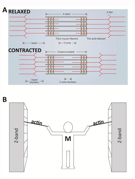
The diagram given below represents the histology of a ... B. I-bands are the light bands and contain actin. C. During muscle contraction the A-band contracts. D. The part between the two Z-lines is called a sarcomere. E. The central part of the thin filament, not overlapped by the thick filament is called H-zone. PDF CHAPTER 9 MUSCLES - Warner Pacific University Sarcomere H zone Thin (actin) filament Thick (myosin) filament Z disc Z disc M line (c) Small part of one myofibril enlarged to show the myofilaments responsible for the banding pattern. Each sarcomere extends from one Z disc to the next. Muscle_Contraction_Student_Version_FORMATTED ... - Course Hero Label the thick and thin filaments in Figs. A, B, and C above. 12. There are three sarcomeres shown in the diagram below. Sarcomere 1 Sarcomere 2 Sarcomere 3 a) In Sarcomere 1, identify the location within the sarcomere of the cross section indicated by Figure A in Model 3. PDF MUSCLE STRUCTURE CHAPTER 5 AND FUNCTION - Weebly C) Tendons function to move various parts of the skeleton in response to skeletal muscle contraction. D) One end of the tendon is usually linked to bone and the other to a skeletal muscle. E) None of the above. Answer: B 2. The contractile unit of a muscle is: A) muscle fibre B) myofilament (myofibril) C) sarcomere D) myosin E) actin Answer: C 3.
Skeletal muscle - Wikipedia
Sarcomere - Definition, Structure, Function and Quiz ... A sarcomere is the functional unit of striated muscle. This means it is the most basic unit that makes up our skeletal muscle. Skeletal muscle is the muscle type that initiates all of our voluntary movement. Herein lies the sarcomere's main purpose. Sarcomeres are able to initiate large, sweeping movement by contracting in unison.

Myogenic Stage, Sarcomere Length, and Protease Activity ...
PDF Muscle Contraction - anatomy & physiology 11. Label the thick and thin filaments in Figs. A, B, and C above. 12. There are three sarcomeres shown in the diagram below. a) In Sarcomere 1, identify the location within the sarcomere of the cross section indicated by Figure A in Model 3. Draw a vertical line and label it A. b) In Sarcomere 2, identify the location within the sarcomere of ...

Site-specific phosphorylation of myosin binding protein-C ...
PDF Muscle Contraction Instructor's Guide - Mr. Jeremy T. Rosen locations within a sarcomere. QUESTIONS: 11. Label the thick and thin filaments in Figs. A, B, and C above. 12. There are three sarcomeres shown in the diagram below. Sarcomere 1 Sarcomere 2 Sarcomere 3 a) In Sarcomere 1, identify the location within the sarcomere of the cross section indicated by Figure A in Model 3.

Cardiac myosin-binding protein C interaction with actin is ...
PDF Internet Activity: Muscle Contractions Read through the ... What needs to be released in order to have the power stroke occur the sarcomere? b. What causes the muscle to contract? ... Click through the first part of the activity to review the muscle structure. Label the diagram below and ... Match the descriptions with the correct muscle fiber structure. Use the words above, and put the term on the line ...
Ultrastructure of Muscle - Skeletal - Sliding Filament ...
Chapter 9 Pictures Flashcards - Quizlet 3) Identify the specific structure indicated by Label G. A) Muscle fascicle B) Triad C) Myofibril D) Sarcomere E) Myofilament

Sarcomere - an overview | ScienceDirect Topics
Solved Thick-filer Muscle Contraction Model 1: Anatomy of ... A Fig. B Fig. QUESTIONS: 10. Label the thick and thin filaments in Figs. A, B, and C above 11. There are three sarcomeres shown in the diagram below. Sarcomere 1 Sarcomere 2 Sarcomere 3 a) In Sarcomere 1, identify the location within the sarcomere of the cross section indicated by Figure A in Model 3.
11.2 Muscles and Movement | BioNinja
PDF The diagram shows part of a muscle myofibril - StudyWise 1. The diagram shows part of a muscle myofibril (a) Name the protein present in the filaments labelled W and X W = myosin, X = actin (b) Figure 2 shows the cut ends of the protein filaments when the myofibril was cut at position Y. figure 3 shows the protein filaments when the myofibril was cut at the same

Muscle tissue - Knowledge @ AMBOSS
Saint Louis Public Schools / Homepage Recognize a sarcomere as a complete contractile unit. Relate the structure of the sarcomere to the distribution of actin and myosin within the myofibril. Describe the sliding-filament hypothesis of muscle contraction. Identify the role of calcium ions (Ca2+), the troponin-tropomyosin complex, and ATP. Recognize that

Given below is the figure of a sarcomere. Identify the parts ...
DOCX (ii) Explain how these structures help in the absorption of substances from the small intestine. (1) (b) (i) The scale bar on this drawing represents a length of 0.1μm.
Sliding Filament Theory, Sarcomere, Muscle Contraction ...
A & P 1 Chapter 10 Test Flashcards | Quizlet Which of the labeled structures on the diagram holds muscles with similar functions together, allows free movement of muscles, and fills spaces between muscles? ... In the figure above, what letter correctly identifies tropomyosin? B. ... which structure helps return a stretched sarcomere to its resting length? C.

Skeletal MyBP-C isoforms tune the molecular contractility of ...
Al-Bana, Haya.Muscle Contraction.Anatomy and... - Course Hero There are three sarcomeres shown in the diagram ft Sarcomere 1 Sarcomere 2 Sarcomere a) In Sarcomere 1, identify the location within the sarcomere of the cross indicated by Figure A in Model 3. Drawa vertical line and at various oo A B o C C above. below. 3 section label it A. 6)
Solved
BSC 2011 undergraduate Flashcards - Quizlet Identify the structures labeled A, B, and C in the diagram of a sarcomere above. A = Actin filament; B = Myosin filament; C = Titin The sliding filament theory states that during muscle contraction

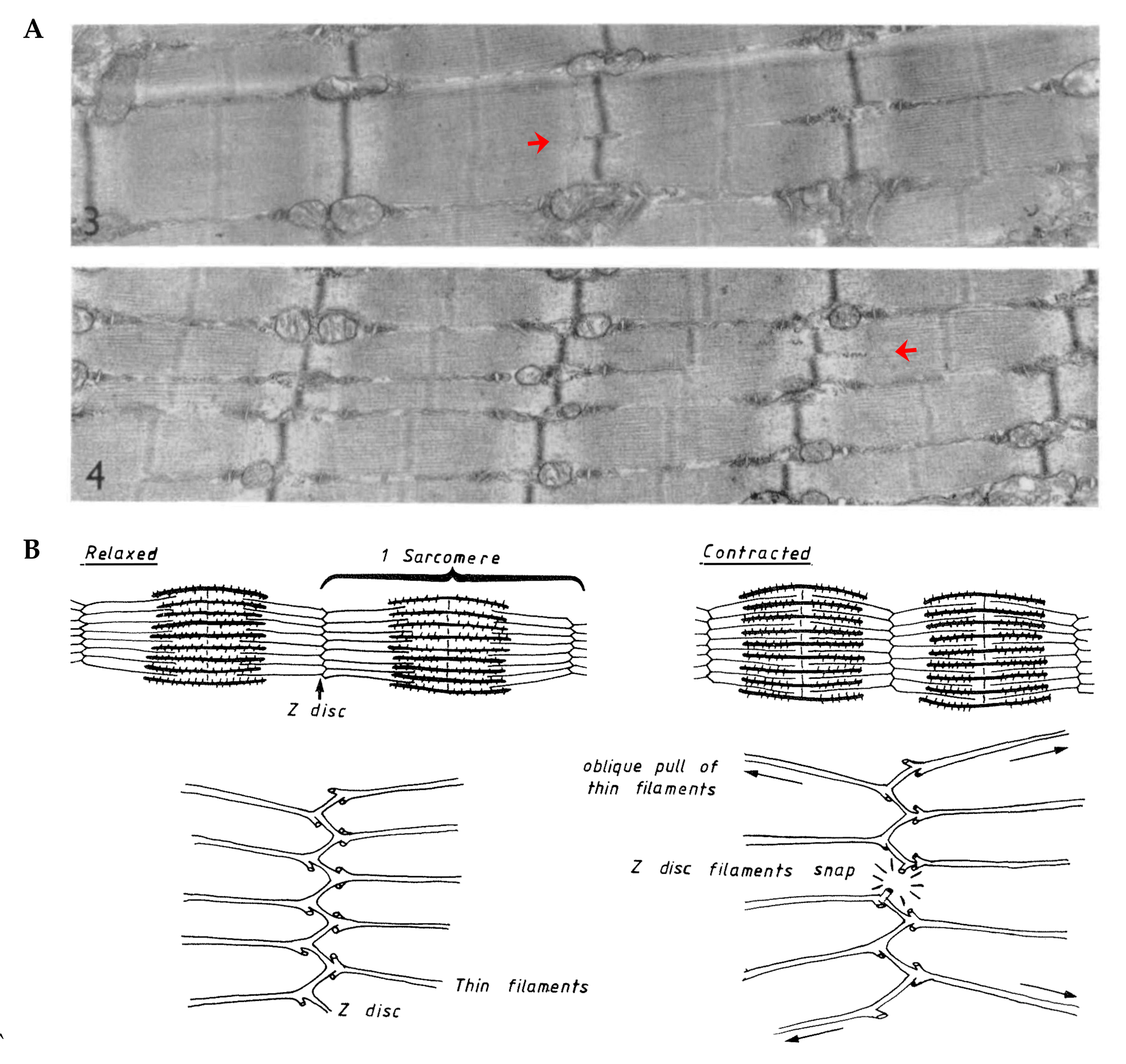
Effects of deleting C-zone repeats on A-band width. a Left ...
Labeled Sarcomere Diagram Jan 23, 2019 · Labeled Sarcomere Diagram. Their observations led to the discovery of sarcomere zones. Sarcomere The figure depicts the structure of a Sarcomere. (Each zone is labeled). They first. Start studying Sarcomere Labeling. Learn vocabulary, terms, and more with flashcards, games, and other study tools.

A&P Final- Ch 10 Flashcards | Chegg.com
Study 50 Terms | Chapter 10: Skeletal... Flashcards | Quizlet B) B C) C D) D E) E. A) A. Identify the letter that indicates the endomysium. A) A B) B C) C D) D E) E. ... connects a muscle to underlying structures through a flat sheet or web. B) consists of a neuron and all the muscle fibers it innervates. ... Which region of the sarcomere does not change in length during contraction? A) A band B) H zone C ...

Muscle tissue - Knowledge @ AMBOSS
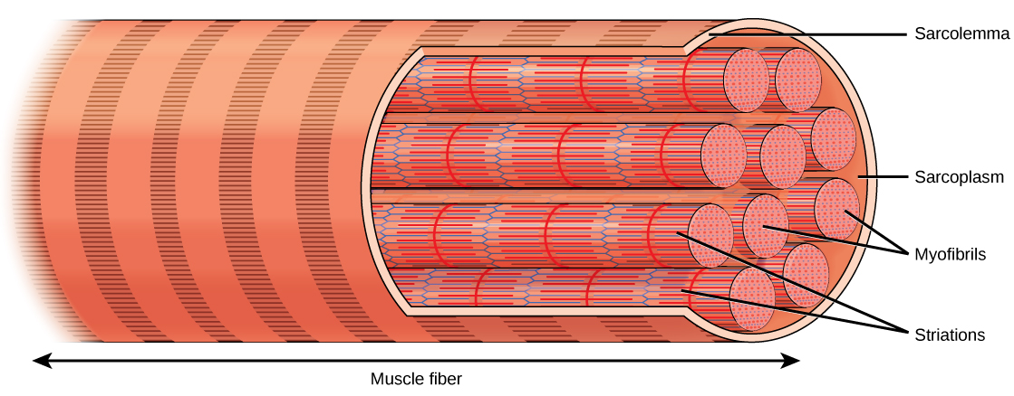
Muscle Cell (Myocyte): Definition, Function & Structure | Biology

Anatomy 10-3 Diagram | Quizlet
Expressing a Z-disk nebulin fragment in nebulin-deficient ...
A)",(B),(C),(D)),("(A-band)",(Z-"line"),(H-"zone"),(I-"band")):}`

Skeletal Muscle Fiber Structures & Sarcomere Flashcards | Quizlet
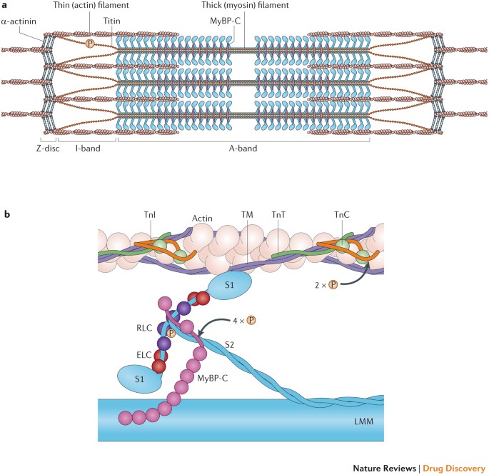
Targeting the sarcomere to correct muscle function | Nature ...

multi choice chapter 10. Muscle Tissue Flashcards - Easy ...

10.2 Skeletal Muscle – Anatomy & Physiology
Cells | Free Full-Text | Identifying the Structural ...

Chapter 9 Homework Flashcards | Quizlet

A) Illustration of skeletal muscle structure copied with ...
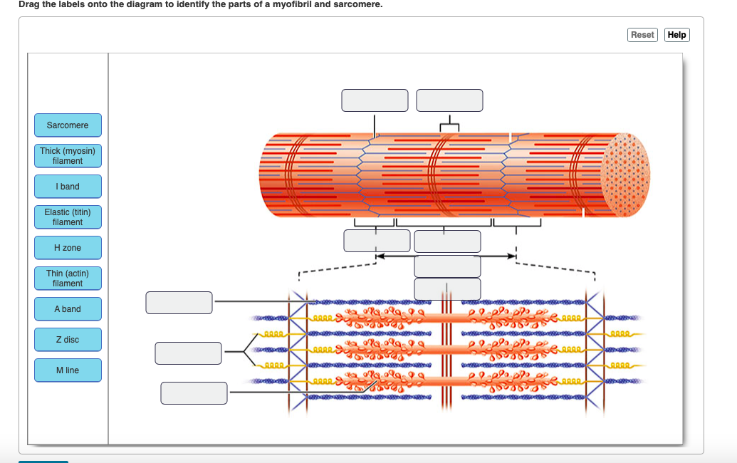
Solved Drag the labels onto the diagram to identify the ...

Mastering A&P Chapter 9 - Muscle and Muscle Tissue Diagram ...

Cardiac Muscle Tissue | Boundless Anatomy and Physiology
multi choice chapter 10. Muscle Tissue Flashcards - Easy ...

UNIT 5: Label the parts of the Sarcomere Flashcards | Quizlet

Study Guide Flashcards | Quizlet

Study Guide Flashcards | Quizlet
IJMS | Free Full-Text | Current Understanding of Residual ...

Muscle structure – muscle under the microscope — Science ...
19.4 Muscle Contraction and Locomotion – Concepts of Biology ...

Ch 13 lab Map, Ch 12 lab map, CH 11 Lab MAP, Ch 10 lab map ...

Solved Question 6 Figure 10.1 Basic skeletal muscle | Chegg.com
0 Response to "38 identify the structures labeled a, b, and c in the diagram of a sarcomere above."
Post a Comment