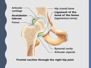
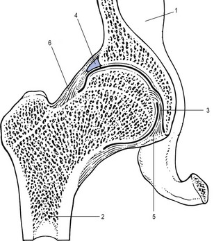
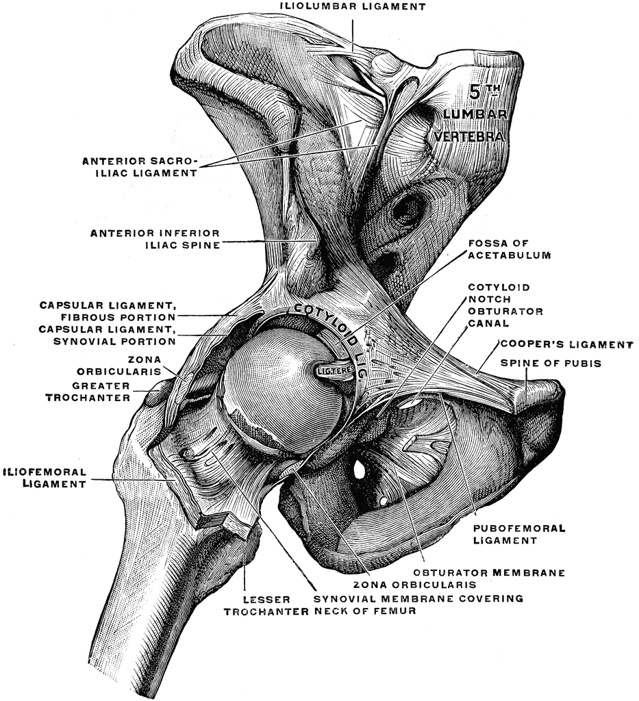
42 the diagram shows a frontal section of the hip joint
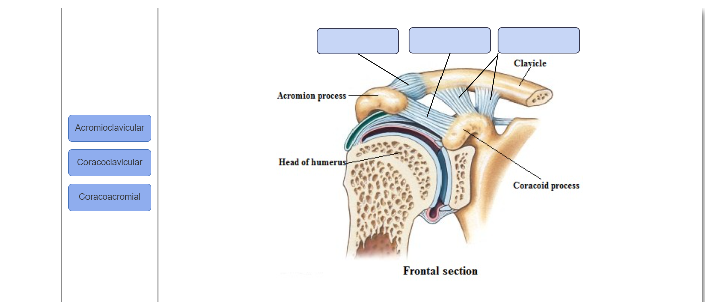
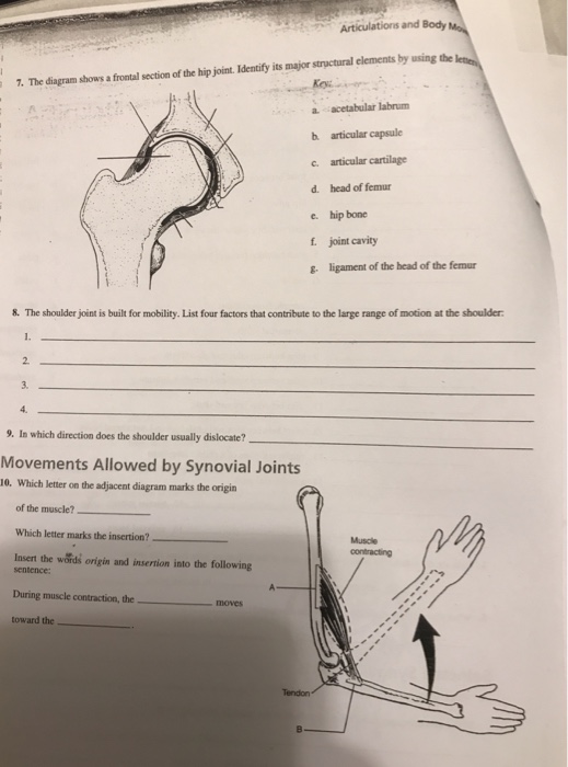
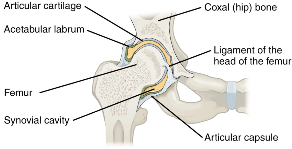
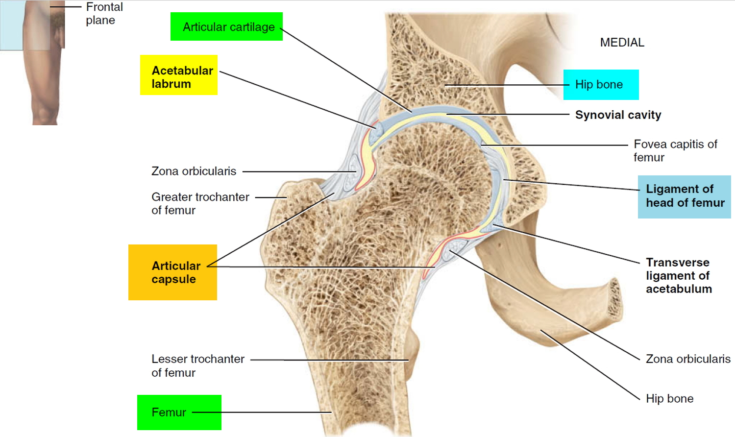
A Labeled Diagram of the Knee With an Insight into Its ... The given diagram of the knee joint can help you to understand its various parts and the description given below will give you an insight of the functioning of the knee. ⚫ Bone There are three bones in the knee namely the femur which is the thigh bone, tibia which is the shin bone and patella which is the knee cap. Joints Flashcards - Quizlet Identify the major structural elements of this frontal section of a hip joint.-acetabular labrum-articular capsule-articular cartilage-coxal bone-head of femur-ligamentum teres-synovial cavity. origin = A insertion = B insertion, origin. Label the origin and insertion points on the diagram below and complete the following statement: During ...
Bones and landmarks of the hip - Musculoskeletal Portfolio The Diagram below shows the posterior apect of the hip clearly visible are the ILIUM and the SACRUM The highest and largest of the 3 bones which comprise the pelvis is the ilium. As the crest reaches the anterior portion, it alters into the anterior superior iliac spine.

The diagram shows a frontal section of the hip joint
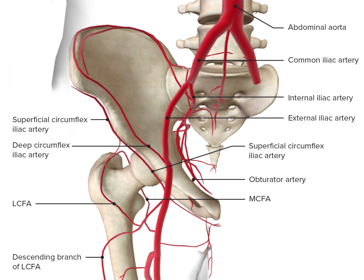
Anatomy of lower extremity - eAnatomy - IMAIOS A diagram shows the various inguinal lymph nodes (lymphatic ganglia). The chapter on the innervation of the lower limb presents diagrams of the lumbosacral plexus and its main nerve branches for the lower limb (lateral cutaneous nerve of the thigh, femoral nerve, sciatic nerve and posterior cutaneous nerve of the thigh and obturator nerve). PDF Cambridge International AS & A Level (b) The diagram shows some stages in a hurdler's technique. A B Identify the items 1-6 in the table to describe a movement analysis of the knee joint and the hip joint of the front/lead (left) leg of the athlete (indicated with a black foot) from position A to position B. Your analysis should include the type of synovial joint, the type of ... A Guide to Hip Anatomy: Bones, Muscles, Tendons & Pain ... The hip is a complicated mechanism and therefore hip pain can originate in many different parts of the joint. Learning the anatomy of your hip will better enable you to pinpoint your pain and work ...
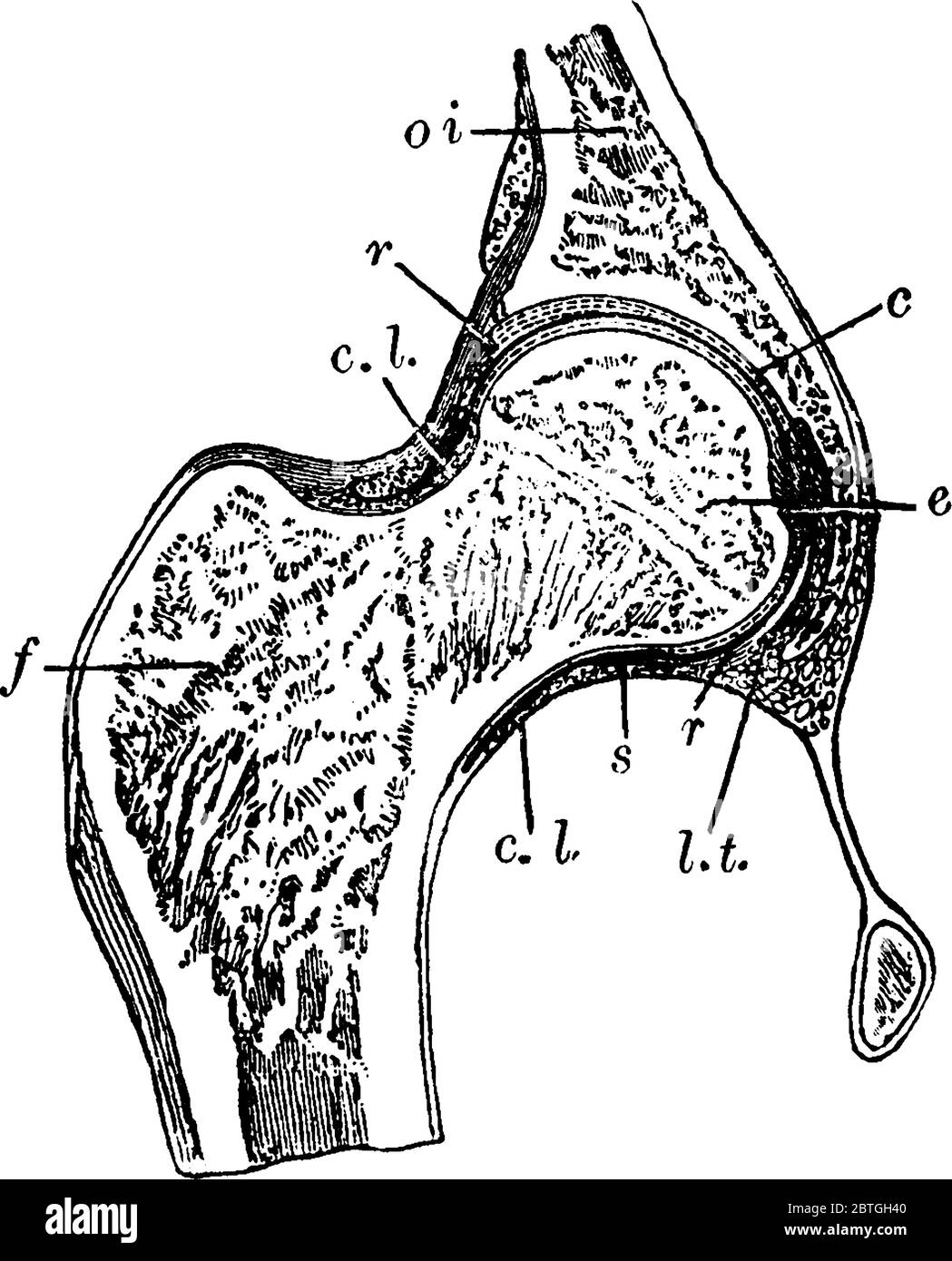
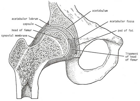
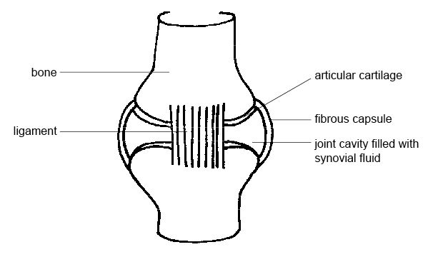
The diagram shows a frontal section of the hip joint. Solved Review Sheet 11 183 7. The diagram shows a frontal ... The diagram shows a frontal section of the hip joint. Identify its major structural elements by using the key letters. Key: a. acetabular labrum b. articular capsule c. articular cartilage d. coxal bone e. head of femur f. ligament of the head of the femur g. synovial cavity 8. The shoulder joint is built for mobility. Hip Anatomy Diagram: From Bones To Joints - Science Trends The hip is a ball-and-socket joint, similar to the joint in the shoulder. Part of the reason for the hip's stability is that there is a very deep socket, called the acetabulum, in the hip joint. A strong capsule joint supported by ligaments and muscles also provides extra stability to the hip. Anatomy & Physiology Ch 9 Flashcards | Quizlet Anatomy & Physiology Ch 9. Which of the following types of joints lacks a joint cavity and is held together by a fibrous connective tissue? 1. Fibrous joints. 2. Cartilaginous joints. 3. Synovial joints. Most of the freely movable joints of the body could be classified both structurally and functionally as __________. Ligaments, tendons, and muscles of the hip joint | Naples ... Ligaments, tendons, and muscles play an important role in the function of the hip. Ligaments are soft tissue structures that connect bones to bones.A joint capsule is a watertight sac that surrounds a joint.In the hip, the joint capsule is formed by a group of three strong ligaments that connect the femoral head to the acetabulum.
Pearson eText15 - Review 7 The diagram shows a frontal ... View Pearson eText15 from BIO 201 at Pima County Community College. Review Sheet 11 183 7. The diagram shows a frontal section of the hip joint. Identify its major structural elements by using the Given diagram shows the bone of the left human hindlimb ... Given diagram shows the bone of the left human hindlimb as seen from the front. ... It is connected to the hip bone by a ball and a socket joint. The femur is a long, curved, robust bone. ... The upper part of the tibia is connected to the joint of the knee, while the lower part is connected to the joint of the ankle. The tibia can be divided ... image.jpg - Articulations and Body Movements 7 The diagram ... View Test Prep - image.jpg from BIOL 2401 at Atascocita H S. Articulations and Body Movements 7. The diagram shows a frontal section of the hip joint. Identify its major structural elements by using Female Pelvis Bones Diagram & Function | Body Maps The pelvis forms the base of the spine as well as the socket of the hip joint. The pelvic bones include the hip bones, sacrum, and coccyx. The hip bones are composed of three sets of bones that ...
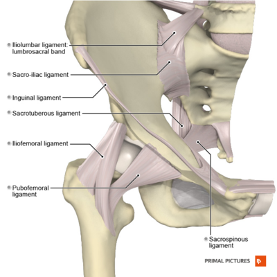
Hip and thigh: Bones, joints, muscles | Kenhub Hip and thigh (posterior view) If you've ever watched the videos for Shakira's Hips don't lie or Justin Timberlake's Can't stop the feeling, you must've wondered how these artists can create such a wide range of hip movements.Well, they have exactly the same anatomy as all of us who use those muscles to support us while we spend countless hours sitting studying the textbooks. Solved > 1.Use key responses to identify the joint types ... 3.Match the synovial joint categories in column B with their descriptions in column A. Column A Column B ... 4.Indicate the number of planes in which each joint can move. uniaxial joints biaxial joints multiaxial joints 5.What characteristics do all joints... 7.The diagram shows a frontal section of the hip joint. Hip joint: Bones, movements, muscles | Kenhub Hip joint (Articulatio coxae) The hip joint is a ball and socket type of synovial joint that connects the pelvic girdle to the lower limb. In this joint, the head of the femur articulates with the acetabulum of the pelvic (hip) bone.. The hip joint is a multiaxial joint and permits a wide range of motion; flexion, extension, abduction, adduction, external rotation, internal rotation and ... PDF Chapter 9 The Hip Joint and Pelvic Girdle - Kean University The Hip Joint and Pelvic Girdle Manual of Structural Kinesiology R.T. Floyd, EdD, ATC, CSCS ... - in frontal plane right pelvis moves inferiorly in relation to left pelvis; either right pelvis rotates downward or left pelvis rotates upward; right lateral tilt ©2007 McGraw-Hill Higher Education.
8 Joints: Part B. - ppt download
Hip Joint - Anatomy Pictures and Information The hip joint is one of the most important joints in the human body. It allows us to walk, run, and jump. It bears our body's weight and the force of the strong muscles of the hip and leg. Yet the hip joint is also one of our most flexible joints and allows a greater range of motion than all other joints in the body except for the shoulder.

Frontal section through the region of pelvic symphyses in the ...
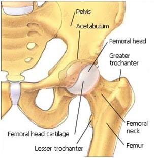
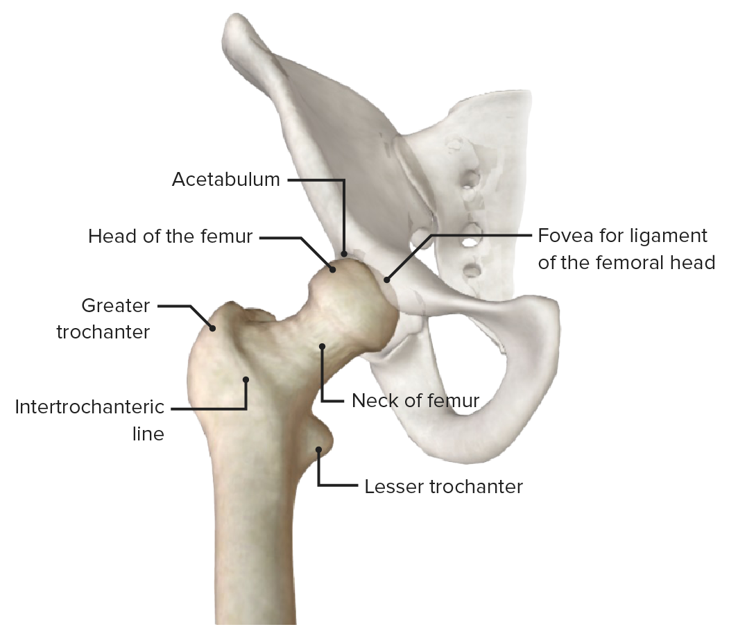
PDF Section 33: Hip - Structural Components on the lateral aspect of the hip bone • Articulates with the head of the femur toArticulates with the head of the femur to form the hip joint • Th Ili I hi d P bi j i t fThe Ilium, Ishium, and Pubis join to form the acetabulum 33-8 From: Howard and Rivera

Acupuncture treats hip pain and sciatica | Jinhee Yoo ...
Chart of Major Muscles on the Front of the Body with Labels This large triangular muscle wraps around the shoulder joint and connects the scapula, clavicle (collar-bone) and humerus. It is a 3-part muscle with anterior (front), middle, and posterior (back) heads. It is controlled by the axillary nerve. The front fibers flex the arm and the middle fibers help abduct the arm (bring the arm away from the ...

Hip & Thigh - Atlas of Anatomy
Solved The diagram shows a frontal section of the hip ... The diagram shows a frontal section of the hip joint. Identity its major structural elements by using the letters Key: a. acetabular labrum b. articular capsule c. articular cartilage d. head of femur e. hip bone f. joint cavity g. ligament of the bead of the femur The shoulder joint is built for mobility.

Pearson eText15 - Review Sheet 11 183 7. The diagram shows a ...
9.4 Synovial Joints - Anatomy & Physiology The joint with the greatest range of motion is the ball-and-socket joint. At these joints, the rounded head of one bone (the ball) fits into the concave articulation (the socket) of the adjacent bone (see Figure 9.4.3f). The hip joint and the glenohumeral (shoulder) joint are the only ball-and-socket joints of the body.

Frontal section through elbow joint vintage Vector Image
What are the Groin Muscles? (with pictures) Shelby Miller Date: March 08, 2022 Groin muscles help support the hip joint.. The groin muscles are a group of muscles situated high on the leg in the inner thigh. This group includes the adductor magnus, adductor longus, and adductor brevis muscles, as well as the pectineus and gracilis.
Joints II
Experiencing Front of Hip Pain? Here's What's Causing It. The diagram on the left is a back view of the hip joint showing the thigh bone (femur) going into the pelvis bone held together by ligaments. The diagram on the right shows a cross section of the hip. As you can see, the top of the femur is shaped like a ball and the concave cavity of the pelvis is shaped like a socket.

Ein text-Buch im allgemeinen Physiologie und Anatomie ...
HCC Learning Web 7. The diagram shows a frontal section of the hip joint. Identify its major structural elements by using the key letters. Key: 185 a. b. C. d. acetabular labrum articular capsule articular cartilage coxal bone head of femur ligamentum teres synovial cavity 8. The shoulder joint is built for mobility.

Cross-sectional view of the normal hip joint. | Download ...
Human Skeletal System | ClipArt ETC Frontal Section Through Hip Joint. Frontal section through the right hip joint, viewed from in front. ... This illustration shows a front view of the human skeleton. Human skeleton. A human skeleton. ... Shown is a a diagram of a diarthrodial joint. In the diarthrodial group the extensive cavity has produced…
Solved Exercise 9 Review Sheet: Articulations 4 of 21 ...
(PDF) Support for total hip replacement surgery ... literature shows a deficiency in studies, thus, the ... only the frontal plane is . considered ... because the socket of the hip joint is part of the pelvis .
Solved The diagram shows a frontal section of the hip joint ...
(Get Answer) - The diagram shows a frontal section of the hip joint ... The diagram shows a frontal section of the hip joint. Identity its major structural elements by using the letters Key: a. acetabular labrum b. articular capsule c. articular cartilage d. head of femur e. hip bone f. joint cavity g. ligament of the bead of the femur The shoulder joint is built for mobility.

Healthy ankle and ankle affected by hemophilic arthropathy ...
A Guide to Hip Anatomy: Bones, Muscles, Tendons & Pain ... The hip is a complicated mechanism and therefore hip pain can originate in many different parts of the joint. Learning the anatomy of your hip will better enable you to pinpoint your pain and work ...
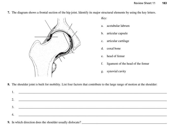
Solved Review Sheet 11 183 7. The diagram shows a frontal ...
PDF Cambridge International AS & A Level (b) The diagram shows some stages in a hurdler's technique. A B Identify the items 1-6 in the table to describe a movement analysis of the knee joint and the hip joint of the front/lead (left) leg of the athlete (indicated with a black foot) from position A to position B. Your analysis should include the type of synovial joint, the type of ...

The Knee Joint Laminated Anatomy Chart
Anatomy of lower extremity - eAnatomy - IMAIOS A diagram shows the various inguinal lymph nodes (lymphatic ganglia). The chapter on the innervation of the lower limb presents diagrams of the lumbosacral plexus and its main nerve branches for the lower limb (lateral cutaneous nerve of the thigh, femoral nerve, sciatic nerve and posterior cutaneous nerve of the thigh and obturator nerve).
Hip Joint Black and White Stock Photos & Images - Alamy
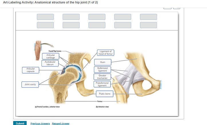
Art-Labeling Activity: Anatomical structure of the | Chegg.com

Diagram of frontal sections of the rabbit's brain showing ...

Part 5 Joints. - ppt video online download
hip joint
Hip Anatomy - Physiopedia

A frontal section of one of the reconstructions shows the ...

Frontal Section Through Hip Joint | ClipArt ETC
The Skeleton Test Yourself Answers - WikiEducator
Anatomy

Hip Pain Explained - including structures & anatomy of the ...
Why does the front of my hip pinch?

Mariam Maged Physiotherapist - DID YOU KNOW? PhysioMM 003 HIP ...

Frontal section through the hip joint (Module 7) Diagram ...

Frontal Section Stock Illustrations – 382 Frontal Section ...
Hip Joint: Anatomy | Concise Medical Knowledge

Regional Biomechanics Hip Joint - ppt video online download

Joints & Joint Movements - ppt video online download
Hip Joint: Anatomy | Concise Medical Knowledge
Hip Dysplasia Baby & Adults - Causes, Symptoms, Surgery ...
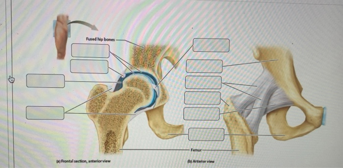
Solved Rused hip bones 10 Femur a) Frontal section, anterior ...
anatomy of the hip and buttock | Musculoskeletal Key

a) Mean time series across subjects at each condition of the ...
Articular Stock Illustrations – 1,437 Articular Stock ...
Hip Joint Seen from Before | ClipArt ETC
Hip Joint: Anatomy | Concise Medical Knowledge

Front hip pain: Causes and treatment

Articulations (joints) - ppt video online download
0 Response to "42 the diagram shows a frontal section of the hip joint"
Post a Comment