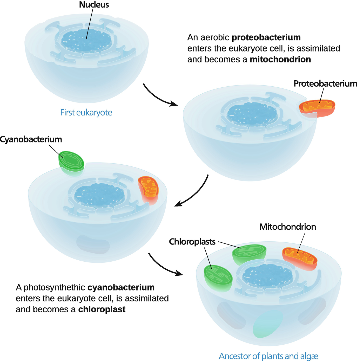
37 drag the labels to the appropriate locations in this diagram.
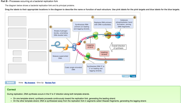
Part A - Animal cell structure Drag the labels onto the diagram to identify the structures of an animal cell. ANSWER: Correct Help Reset Help Reset Smooth endoplasmic reticulum (ER) Central vacuole Nucleus Cell wall Mitochondrion Rough endoplasmic reticulum (ER) Chloroplast Golgi apparatus Cytosol Cytoskeleton Golgi Apparatus Mitochondrion Nucleus Plasma Membrane Ribosomes Rough Endoplasmic Reticulum (ER) Smooth Endoplasmic Reticulum (ER) Drag the labels to their appropriate locations in the diagram to describe the name or function of each structure. Use pink labels for the pink targets and blue labels for the blue targets. A) Breaks hydrogen bonds, unwinding DNA double helix. B) Synthesizes RNA primers on leading and lagging strands. C) Replaces RNA primers with DNA nucleotides.
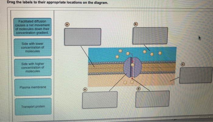
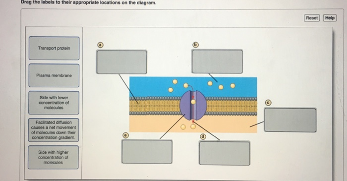
18. Label the following diagram: Part A - Diffusion Drag the labels to their appropriate locations on the diagram. Side with higher concentration of molecules (b) Plasma membrane Side with lower concentration of molecules Diffusion causes a net movement of molecules down their concentration gradient. check_circle.
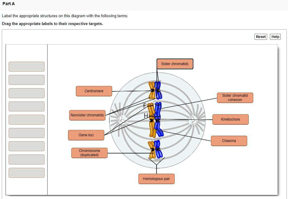
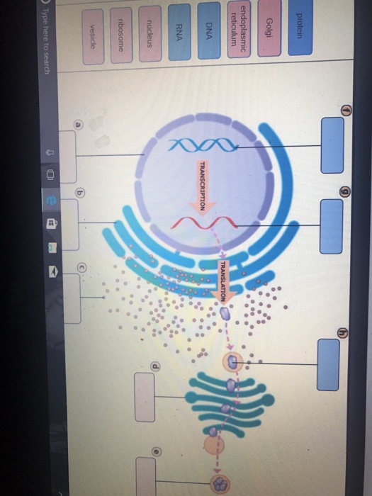
Drag the labels to the appropriate locations in this diagram.
Drag the labels to their appropriate locations in the flowchart below, indicating the sequence of events in the production of fragment B. (Note that pol I stands for DNA polymerase I, and pol III stands for DNA polymerase III.) step 2 after pol III binds to 3' end of primer B. pol I binds to 5' end of primer A. 17/11/2021 · Home › 41 drag the labels onto the diagram to identify the steps in a reaction both with and without enzymes. 41 drag the labels onto the diagram to identify the steps in a reaction both with and without enzymes. Written By Eric Y. Gallegos Wednesday, November 17, 2021 Add Comment Edit. The answer depends on the enzyme. Drag the correct label under each diagram. Left: exocytosis - a process in which material inside a cell is packaged into vesicles and excreted into the extracellular medium Right: endocytosis - a process in which the plasma membrane invaginates or fold inward, to form a vesicle that brings substances into the cell
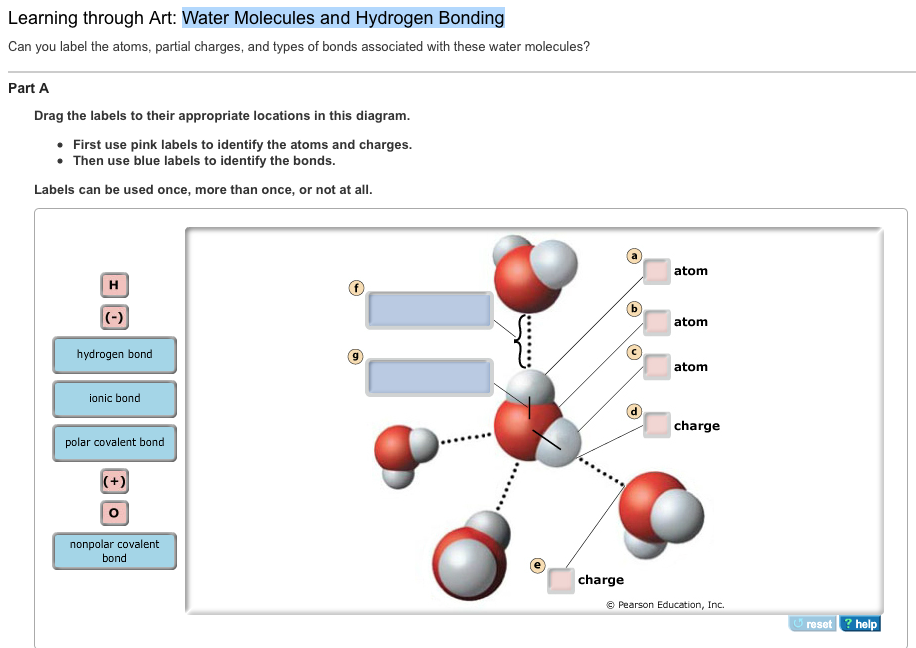
Drag the labels to the appropriate locations in this diagram.. Drag The Correct Label To The Appropriate Location To Identify The Parts Of An Osteon. This problem has been solved. Part a homologous chromosomes drag the labels onto the diagram to identify the various chromosome structures. Drag the labels from the left to their correct locations in the concept map on the right. Part A - Hydrogen bonding Label the following diagram of water molecules, indicating the location of bonds and the partial charges on the atoms. Drag the labels to their appropriate locations on the diagram of the water molecules below. Labels can be used once, more than once, or not at all. Hint 1. Drag the labels to their appropriate locations on the diagram of the water molecules below. Labels can be used once, more than once, or not at all. Hint 1. Electronegativity and polar covalent bonds In covalent bonds, the electrons are not always shared equally between the atoms. Some atoms hold electrons more tightly than others. The diagram below shows a replication fork with the two parental DNA strands labeled at their 3' and 5 4/4 (5). The diagram below shows a bacterial replication fork and its principal proteins. Drag the labels to their appropriate locations in the diagram to describe the name or function of each structure. Use pink labels for the pink targets ...
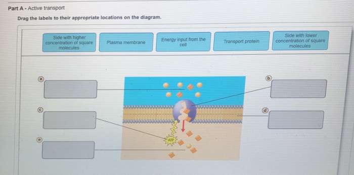
Drag the labels to their appropriate locations on the diagram. a. side with lower concentration of square molecules b. transport protein c. energy input from the cell d. plasma membrane e. side with higher concentration of square molecules. Some large molecules move into or out of cells by exocytosis or endocytosis. Part A. Active Transport. Drag the labels to their appropriate locations on the diagram. Learn this topic by watching Transporters Concept Videos. Drag the correct labels to the appropriate locations in the diagram to show the composition of the daughter DNA molecules after one and two cycles of DNA replication. In the labels, the original parental DNA is blue and the DNA synthesized during replication is red. Drag the labels to their appropriate locations to complete the Punnett square for Morgan's F1 x F1 cross. Drag pink labels onto the pink targets to indicate the alleles carried by the gametes (sperm and egg). Drag blue labels onto the blue targets to indicate the possible genotypes of the offspring.
Drag the labels to their appropriate locations on the diagram. Learn this topic by watching Passive Transport: Diffusion and Osmosis Concept Videos All Cell Biology Practice Problems Passive Transport: Diffusion and Osmosis Practice Problems Drag the labels to their appropriate locations in this diagram. First use the labels of Group 1 to identify the atoms and charges. Then use the labels of Group 2 to identify the bonds. Labels can be used once, more than once, or not at all. Keys: (-) (+) H O Ionic Bond Polar covalent bond Hydrogen Bond nonpolar covalent bond Part A - Regulating blood sugar This diagram shows how the body keeps blood glucose at a normal level. Drag each label to the appropriate location on the diagram. ANSWER: Correct BioFlix Quiz: Homeostasis: Regulating Blood Sugar Watch the animation then answer the questions. Part A When blood glucose levels are high Hint 1. The diagram below shows a bacterial replication fork and its principal proteins. Drag the labels to their appropriate locations in the diagram to describe the name or function of each structure. Use pink labels for the pink targets and blue labels for the blue targets. A)breaking hydrogen bonds, unwinding DNA double helix ...
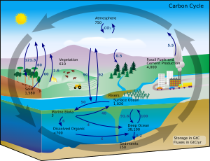
View full document. See Page 1. Drag the labels to their appropriate locations on the diagram of the carbon cycle. First, drag the blue labels to the blue targets to identify the reservoirs in the carbon cycle. Then drag the pink labels to the pink targets to identify the processes in the carbon cycle. Submit Hints My Answers Give Up Review Part.
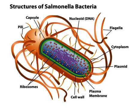
Drag the labels to the appropriate locations in this diagr… Get the answers you need, now! seanders76 seanders76 02/13/2021 Biology College answered • expert verified Can you label the structures of a prokaryotic cell? Drag the labels to the appropriate locations in this diagram 2 See answers Advertisement Advertisement
Drag the labels to their appropriate locations on the diagram below. Targets of Group 1 can be used more than once. Hint 1. Distinguishing the 3' and 5' ends of a DNA strand A strand of DNA consists of a linear polymer of DNA nucleotides.
Certain molecules use diffusion to cross the plasma membrane. Drag the labels to their appropriate locations on the diagram. a. side with higher concentration of molecules. b. plasma membrane. c. side with lower concentration of molecules. d. diffusion causes a net movement of molecules down their concentration gradient.
Drag the labels to the appropriate locations on the diagram of the thylakoid membrane. Use only the blue labels for the blue targets, and only the pink labels for the pink targets. Note: One blue target and one pink target should be left empty.
Drag the correct label under each diagram. Left: exocytosis - a process in which material inside a cell is packaged into vesicles and excreted into the extracellular medium Right: endocytosis - a process in which the plasma membrane invaginates or fold inward, to form a vesicle that brings substances into the cell
17/11/2021 · Home › 41 drag the labels onto the diagram to identify the steps in a reaction both with and without enzymes. 41 drag the labels onto the diagram to identify the steps in a reaction both with and without enzymes. Written By Eric Y. Gallegos Wednesday, November 17, 2021 Add Comment Edit. The answer depends on the enzyme.
Drag the labels to their appropriate locations in the flowchart below, indicating the sequence of events in the production of fragment B. (Note that pol I stands for DNA polymerase I, and pol III stands for DNA polymerase III.) step 2 after pol III binds to 3' end of primer B. pol I binds to 5' end of primer A.







![Expert Answer] Drag labels to the appropriate locations in ...](https://us-static.z-dn.net/files/dd9/363dc6f0791b99410cbee879fdbd9356.png)








0 Response to "37 drag the labels to the appropriate locations in this diagram."
Post a Comment