37 wiggers diagram aortic regurgitation
27. Can we use the term "afterload" for aortic pressure during the phase that the aortic valve is closed? _____ _____ 28. Above diagram shows the relationship of Frank-Starling curves to afterload and stroke volume. The centrally located curve marked "B" is for normal heart. Which of the two curves; A or C, represents increased and ... The Wright table is introduced as a novel tool for teaching and learning the cardiac cycle. It supplements the nearly 100-yr-old Wiggers diagram, which is information rich but difficult for many students to learn. The Wright table offers a compact presentation of information, viewable both in terms of how 1) each compartment's pressures and flows change over time; and 2) the heart works as a ...
Early diastolic murmurs immediately follow S2. Examples: aortic and pulmonary regurgitation. Mid-diastolic murmurs (rumble) are due to increased flow (relative stenosis) through the mitral (VSD) or the tricuspid valves (ASD).; Late diastolic murmurs are due to pathological narrowing of the atrioventricular (AV) valves. Example: rheumatic mitral stenosis. Tricuspid stenosis is very rare in childr

Wiggers diagram aortic regurgitation
Aortic regurgitation occurs when the aortic valve fails to close completely and blood flows back from the aorta (Ao) into the left ventricle after ejection into the aorta is complete and during the time that the left ventricle (LV) is also being filled from the left atrium (LA) (see figure at right). Because the ventricle is being filled from two sources (aorta and LA), this leads to much ... Aortic valve stenosis is characterized by the left ventricular pressure being much greater than aortic pressure during left ventricular (LV) ejection (see figure at right). In this example, LV peak systolic pressure during ejection is 200 mmHg (normally ~120 mmHg) and the aortic pressure is slightly reduced to from 120 to 110 mmHg. position of aortic valve during ventricular filling due to high ventricular pressure. ... changes in Wiggers diagram R heart compared to L heart. all pressures would be lower, valve opening and ventricular volumes would be the same ... regurgitation. murmur often heard throughout systole over the right AV valve.
Wiggers diagram aortic regurgitation. Wigger's Diagram demonstrating the physiology of diastolic mitral regurgitation. The diagram shows the rapid decrease in diastolic aortic pressure and increase in left ventricular pressure caused by severe aortic regurgitation. FURTHER OBSERVATIONS ON EXPERIMENTAL AORTIC INSUFFICIENCY II. CINEMATOGRAPHIC STUDIES OF CHANGES IN VENTRICULAR SIZE AND IN LEFT VENTRICULAR DISCHARGE By CARLJ. WIGGERS, HAROLD THEISENAND HARLEYA. WILLIAMS (FromtheDepartmentofPhysiology, WesternReserve University MedicalSchool, Clevdand) (Receivedforpublication April30, 1930) INTRODUCTION Aortic regurgitation. Aortic regurgitation (AR) occurs when there is backflow of blood from the aorta into the left ventricle during ventricular diastole. Aortic regurgitation is associated with an early diastolic murmur heard loudest at the left sternal edge Aetiology. Aortic regurgitation can be either acute or chronic. Chronic AR is often ... Wigger's diagram: This seemingly complex diagram (Don't Panic!) is actually a very useful tool to learn the components of the cardiac cycle and how they relate to cardiac exam findings, which I have indicated in blue. To start, identify the aortic, LV and LA pressure lines and talk yourself through the sequence of
The "Wiggers diagram" ... Mitral and aortic regurgitation are the most common regurgitant lesions. Alternatively, the orifice of a valve may become narrowed or stenotic. This obstructs the flow of blood through it and requires increased pressure gradients to be generated across the valve to achieve Aortic/Pulmonary artery pressure increases slightly during this phase due to blood pushin back against the now closed aortic/pulmonary valve. Figure 2 : (a) The Wigger's diagram, which shows changes in pressure and volume of heart compartments, as well as electrical and auditory signals that can be detected at different stages of the cardiac ... Figure 4.1 Schematic of aortic pressure. Figure 4.2 Wiggers diagram [LVEDV = Left Ventricular End Diastolic Volume; LVESV = Left Ventricular End Systolic Volume]. Figure 4.3 Aortic pressure in a hypovolemic patient. Aortic pressure tracings taken in a patient before and after an intravenous (IV) fluid bolus. Chart and Diagram Slides for PowerPoint - Beautifully designed chart and diagram s for PowerPoint with visually stunning graphics and animation effects. Our new CrystalGraphics Chart and Diagram Slides for PowerPoint is a collection of over 1000 impressively designed data-driven chart and editable diagram s guaranteed to impress any audience.
findings of aortic insufficiency consisting of a wide .. Diagram of the ascending and descending aorta illustrating . Stewart10 and later Wiggers and Green Aortic insufficiency (AI), also known as aortic regurgitation (AR), is the leaking of the aortic valve of the heart that causes blood to flow in the reverse direction. If a high-pressure jet resulting from aortic stenosis is present, the aorta will stretch If we go back to Part I and look at the Wigger's diagram, the aortic valve is. Dr. Carl Wiggers' careful observations have provided a meaningful resource for students to learn how the heart works. Throughout the many years from his initial .Valvular Stenosis. The first set of questions deals with the the events during each of the phases or periods of the normal cardiac cycle. Please use the Wiggers diagrams below showing systemic arterial, left ventricular, and left atrial pressures to answer the questions. In some cases, more than one letter may be circled to achieve the correct answer. What elements of the wigger diagram will look different in mitral regurgitation large v wave and rapid y descent A patient is complaining of trouble with breathing - you auscultate and hear a murmur occurring throughout systole, turbulent flow during diastole, and S3.

Wiggers Diagram Aortic Regurgitation
midsystolic clicks and the mitral regurgitation heard with mitral valve prolapse o Diastolic Murmurs occur between S2 and S1 Early diastolic murmurs: decrescendo murmurs, arise from aortic or pulmonary valve regurgitation Mid-diastolic murmurs: diamond shaped murmurs, associated with increased

17 Best images about RCIS - waveforms on Pinterest | Cars ...
The cardiac cycle is a series of pressure changes that take place within the heart. These pressure changes result in the movement of blood through different chambers of the heart and the body as a whole. These pressure changes originate as conductive electrochemical changes within the myocardium that result in the concentric contraction of cardiac muscle.


Closeup of skeleton hand model
Left Ventricle Pressure Volume Loop. Wiggers' Diagram. Jugular Venous Pressure. Aortic Stenosis. Aortic Regurgitation. Mitral Stenosis. Mitral Regurgitation. Cor Pulmonale. Heart Failure.

Cardiovascular | themedicallyinclined
Read more: Aortic Regurgitation in Chapter 283: Aortic Valve Disease, Harrison's Principles of Internal Medicine 19e. The above Wiggers diagrams are modifications of: adh30 revised work by DanielChangMD who revised original work of DestinyQx; Redrawn as SVG by xavax - Wikimedia Commons: Wiggers Diagram.svg, CC BY-SA 4.0 « What causes alcohol withdrawal seizures?

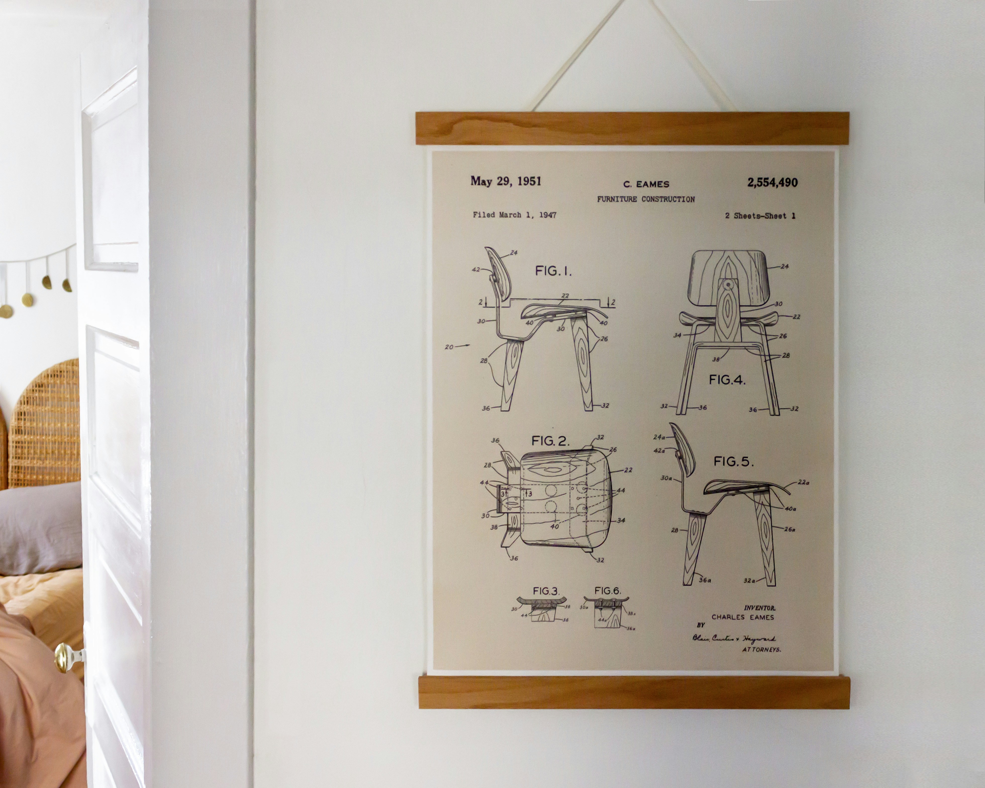
Eames Chair Diagram Schematics - 1951 Chair Poster Hanging on Wall
The QRS wave, on the electrocardiogram section of the Wiggers Diagram (image 5.1) represents the beginning of systole. This phase represents ventricular qi depolarization. The rise in pressure in the ventricles exceeds the pressure in the atrium, causing the AV valves to close. The S1 (first heart sound) is the sound heard during this phase. 2.

S06E04
In early ventricular systole. The semilunar valves open. Ventricular Diastole represents. The period of time the ventricles are filling. Between the end of the T wave and the onset of the QRS complex. Ventricular Diastole. Approximately 70% of the Diastolic filling occurs during. Early Ventricular Diastole.

413 best images about Echocardiography on Pinterest ...
This is well-illustrated on a Wiggers diagram where the QRS complex on the ECG directly precedes ventricular systole (represented on the diagram by increased ventricular pressure). Function ... For example, a murmur determined to be systolic rules out diastolic murmur causes such as mitral stenosis and aortic regurgitation. As a general rule ...


Female civil engineer designs theme park attractions
About Press Copyright Contact us Creators Advertise Developers Terms Privacy Policy & Safety How YouTube works Test new features Press Copyright Contact us Creators ...


Closeup of skeleton foot model
Left Ventricle Pressure Volume Loop. Myocardial Infarction. Ischemic EKG

Isovolumic relaxation time - Wikipedia
Wigger Diagram Set-up: Along the bottom, indicate that we'll track time in seconds from 0 to 0.8, which is the duration of a typical healthy cardiac cycle. At the top of the graph, show that we'll track events of the left atrium and left ventricle, as follows: - The left atrium is in systole from time 0 - 0.1, then enters diastole.

MCAT Memoranda
Aortic regurgitation Aortic regurgitation Aortic insufficiency (AI) is a condition in which the aortic valve fails to close completely at the end of systolic ejection, causing leakage of blood back through the valve during LV diastole. Cardiac Cycle - Summary and Wigger's Diagram. Aortic valve stenosis
")
Stock market chart value. Made with analog vintage lens, Leica APO Macro Elmarit-R 2.8 100mm (Year: 1993)
position of aortic valve during ventricular filling due to high ventricular pressure. ... changes in Wiggers diagram R heart compared to L heart. all pressures would be lower, valve opening and ventricular volumes would be the same ... regurgitation. murmur often heard throughout systole over the right AV valve.

S06E04
Aortic valve stenosis is characterized by the left ventricular pressure being much greater than aortic pressure during left ventricular (LV) ejection (see figure at right). In this example, LV peak systolic pressure during ejection is 200 mmHg (normally ~120 mmHg) and the aortic pressure is slightly reduced to from 120 to 110 mmHg.

17 Best images about RCIS - waveforms on Pinterest | Cars ...
Aortic regurgitation occurs when the aortic valve fails to close completely and blood flows back from the aorta (Ao) into the left ventricle after ejection into the aorta is complete and during the time that the left ventricle (LV) is also being filled from the left atrium (LA) (see figure at right). Because the ventricle is being filled from two sources (aorta and LA), this leads to much ...

Chapter 14 - Cardiovascular Physiology at Nova ...

Aortic Regurgitation

413 best images about Echocardiography on Pinterest ...

Wiggers Diagram Aortic Stenosis

S06E04

Valvular heart disease kay johnstone

Wiggers Diagram Explained | LV Systolic Function ...

S10E03

Cardiovascular 7 & 8: Valvular Heart Diseases flashcards ...

Wiggers Diagram | nursing info/education | Pinterest ...

29 best RCIS - waveforms images on Pinterest | Cardiology ...

Cardiopulmonology | themedicallyinclined


place to be

S3 Week 2 Pathology Flashcards - Cram.com

Physiologic and pathophysiologic function of the heart

S06E04

S10E03

Schneid Guide to Effects of Heart Valve Lesions on the ...

S10E03
.png "MedicoNotebook: PaQ1c2v3 - Sequential Cardiac Cycle events")
MedicoNotebook: PaQ1c2v3 - Sequential Cardiac Cycle events

S10E03


Closeup of skeleton hand model

Medical diagram
0 Response to "37 wiggers diagram aortic regurgitation"
Post a Comment